 Movie
Movie Controller

[English] 日本語
 Yorodumi
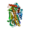
- PDB-3sg1: 2.6 Angstrom Crystal Structure of UDP-N-acetylglucosamine 1-carbo... -
+ Open data

- Basic information
| Entry | Database: PDB / ID: 3sg1 | ||||||
|---|---|---|---|---|---|---|---|
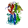
| Title | 2.6 Angstrom Crystal Structure of UDP-N-acetylglucosamine 1-carboxyvinyltransferase 1 (MurA1) from Bacillus anthracis | ||||||
 Components Components | UDP-N-acetylglucosamine 1-carboxyvinyltransferase 1 | ||||||
 Keywords Keywords | TRANSFERASE / Structural Genomics / Center for Structural Genomics of Infectious Diseases / CSGID / Cell wall formation | ||||||
| Function / homology |  Function and homology information Function and homology informationUDP-N-acetylglucosamine 1-carboxyvinyltransferase activity / UDP-N-acetylgalactosamine biosynthetic process / UDP-N-acetylglucosamine 1-carboxyvinyltransferase / peptidoglycan biosynthetic process / cell wall organization / regulation of cell shape / cell division / cytoplasm Similarity search - Function | ||||||
| Biological species |  | ||||||
| Method |  X-RAY DIFFRACTION / SYNCHROTRON / MOLECULAR REPLACEMENT / Resolution: 2.6 Å X-RAY DIFFRACTION / SYNCHROTRON / MOLECULAR REPLACEMENT / Resolution: 2.6 Å | ||||||
 Authors Authors | Minasov, G. / Halavaty, A. / Filippova, E.V. / Shuvalova, L. / Dubrovska, I. / Winsor, J. / Papazisi, L. / Anderson, W.F. / Center for Structural Genomics of Infectious Diseases (CSGID) | ||||||
 Citation Citation | Journal: TO BE PUBLISHED Title: 2.6 Angstrom Crystal Structure of UDP-N-acetylglucosamine 1-carboxyvinyltransferase 1 (MurA1) from Bacillus anthracis. Authors: Minasov, G. / Halavaty, A. / Filippova, E.V. / Shuvalova, L. / Dubrovska, I. / Winsor, J. / Papazisi, L. / Anderson, W.F. / Center for Structural Genomics of Infectious Diseases (CSGID) | ||||||
| History |
|
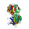
- Structure visualization
| Structure viewer | Molecule: MolmilJmol/JSmol |
|---|
- Downloads & links
-Download
| PDBx/mmCIF format | 3sg1.cif.gz | 642.5 KB | Display | PDBx/mmCIF format |
|---|---|---|---|---|
| PDB format | pdb3sg1.ent.gz | 531.3 KB | Display | PDB format |
| PDBx/mmJSON format | 3sg1.json.gz | Tree view | PDBx/mmJSON format | |
| Others |  Other downloads Other downloads |
-Validation report
| Arichive directory | https://data.pdbj.org/pub/pdb/validation_reports/sg/3sg1ftp://data.pdbj.org/pub/pdb/validation_reports/sg/3sg1 | HTTPS FTP |
|---|
-Related structure data
| Related structure data | 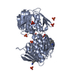 1ejdS S: Starting model for refinement |
|---|---|
| Similar structure data | |
| Other databases |


-Links
 PDBj
PDBj
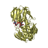
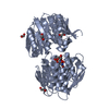
- Assembly
| Deposited unit | 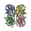
| ||||||||||||||||||||||||||||||
|---|---|---|---|---|---|---|---|---|---|---|---|---|---|---|---|---|---|---|---|---|---|---|---|---|---|---|---|---|---|---|---|
| 1 | 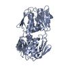
| ||||||||||||||||||||||||||||||
| 2 | 
| ||||||||||||||||||||||||||||||
| 3 | 
| ||||||||||||||||||||||||||||||
| 4 | 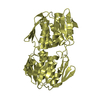
| ||||||||||||||||||||||||||||||
| 5 |
| ||||||||||||||||||||||||||||||
| Unit cell |
| ||||||||||||||||||||||||||||||
| Noncrystallographic symmetry (NCS) | NCS domain:
NCS domain segments: Component-ID: 1 / Ens-ID: 1 / Beg auth comp-ID: MET / Beg label comp-ID: MET / End auth comp-ID: GLU / End label comp-ID: GLU / Refine code: 4 / Auth seq-ID: 1 - 419 / Label seq-ID: 25 - 443
|
-Components
| #1: Protein | Mass: 49573.711 Da / Num. of mol.: 4 Source method: isolated from a genetically manipulated source Source: (gene. exp.) References: UniProt: Q81K13, UDP-N-acetylglucosamine 1-carboxyvinyltransferase #2: Chemical | ChemComp-PG4 / |   Mass: 194.226 Da / Num. of mol.: 1 / Source method: obtained synthetically / Formula: C8H18O5 / Comment: precipitant*YM Mass: 194.226 Da / Num. of mol.: 1 / Source method: obtained synthetically / Formula: C8H18O5 / Comment: precipitant*YM#3: Chemical | ChemComp-PGE / |   Mass: 150.173 Da / Num. of mol.: 1 / Source method: obtained synthetically / Formula: C6H14O4 Mass: 150.173 Da / Num. of mol.: 1 / Source method: obtained synthetically / Formula: C6H14O4#4: Water | ChemComp-HOH / |  Mass: 18.015 Da / Num. of mol.: 494 / Source method: isolated from a natural source / Formula: H2O Mass: 18.015 Da / Num. of mol.: 494 / Source method: isolated from a natural source / Formula: H2O |
|---|
-Experimental details
-Experiment
| Experiment | Method: X-RAY DIFFRACTION / Number of used crystals: 1 |
|---|
- Sample preparation
| Crystal | Density Matthews: 1.9 Å3/Da / Density % sol: 35.26 % |
|---|---|
| Crystal grow | Temperature: 295 K / Method: vapor diffusion, sitting drop / pH: 8.5 Details: Protein solution: 7.7 mg/mL, 0.25M Sodium chloride, 0.01M Tris-HCl (pH 8.3), Screen solution: PEG's II, condition D10, 0.2M Sodium acetate, 0.1M Tris (pH 8.5), 30% (w/v) PEG 4000, VAPOR ...Details: Protein solution: 7.7 mg/mL, 0.25M Sodium chloride, 0.01M Tris-HCl (pH 8.3), Screen solution: PEG's II, condition D10, 0.2M Sodium acetate, 0.1M Tris (pH 8.5), 30% (w/v) PEG 4000, VAPOR DIFFUSION, SITTING DROP, temperature 295K |
-Data collection
| Diffraction | Mean temperature: 100 K |
|---|---|
| Diffraction source | Source: SYNCHROTRON / Site: APS  / Beamline: 21-ID-G / Wavelength: 0.97856 Å / Beamline: 21-ID-G / Wavelength: 0.97856 Å |
| Detector | Type: MARMOSAIC 300 mm CCD / Detector: CCD / Date: Jul 7, 2009 / Details: Beryllium lenses |
| Radiation | Monochromator: Diamond / Protocol: SINGLE WAVELENGTH / Monochromatic (M) / Laue (L): M / Scattering type: x-ray |
| Radiation wavelength | Wavelength: 0.97856 Å / Relative weight: 1 |
| Reflection | Resolution: 2.6→30 Å / Num. all: 45804 / Num. obs: 45804 / % possible obs: 100 % / Observed criterion σ(I): -3 / Redundancy: 3.8 % / Biso Wilson estimate: 45.4 Å2 / Rmerge(I) obs: 0.103 / Net I/σ(I): 12.6 |
| Reflection shell | Resolution: 2.6→2.64 Å / Redundancy: 3.8 % / Rmerge(I) obs: 0.509 / Mean I/σ(I) obs: 2.7 / Num. unique all: 2277 / % possible all: 100 |
- Processing
| Software |
| |||||||||||||||||||||||||||||||||||||||||||||||||||||||||||||||||||||||||||||||||||||||||||||||||||||||||||||||||||||||||||||||||||||||||||||||||||||||||||||||||||||||||||||||||||||||||||||||||||||||||||||||||||||||||||||||||
|---|---|---|---|---|---|---|---|---|---|---|---|---|---|---|---|---|---|---|---|---|---|---|---|---|---|---|---|---|---|---|---|---|---|---|---|---|---|---|---|---|---|---|---|---|---|---|---|---|---|---|---|---|---|---|---|---|---|---|---|---|---|---|---|---|---|---|---|---|---|---|---|---|---|---|---|---|---|---|---|---|---|---|---|---|---|---|---|---|---|---|---|---|---|---|---|---|---|---|---|---|---|---|---|---|---|---|---|---|---|---|---|---|---|---|---|---|---|---|---|---|---|---|---|---|---|---|---|---|---|---|---|---|---|---|---|---|---|---|---|---|---|---|---|---|---|---|---|---|---|---|---|---|---|---|---|---|---|---|---|---|---|---|---|---|---|---|---|---|---|---|---|---|---|---|---|---|---|---|---|---|---|---|---|---|---|---|---|---|---|---|---|---|---|---|---|---|---|---|---|---|---|---|---|---|---|---|---|---|---|---|---|---|---|---|---|---|---|---|---|---|---|---|---|---|---|---|
| Refinement | Method to determine structure: MOLECULAR REPLACEMENT Starting model: 1EJD Resolution: 2.6→29.88 Å / Cor.coef. Fo:Fc: 0.947 / Cor.coef. Fo:Fc free: 0.884 / SU B: 27.754 / SU ML: 0.268 / Isotropic thermal model: Individually Refined / Cross valid method: THROUGHOUT / ESU R Free: 0.373 / Stereochemistry target values: MAXIMUM LIKELIHOOD / Details: HYDROGENS HAVE BEEN ADDED IN THE RIDING POSITIONS
| |||||||||||||||||||||||||||||||||||||||||||||||||||||||||||||||||||||||||||||||||||||||||||||||||||||||||||||||||||||||||||||||||||||||||||||||||||||||||||||||||||||||||||||||||||||||||||||||||||||||||||||||||||||||||||||||||
| Solvent computation | Ion probe radii: 0.8 Å / Shrinkage radii: 0.8 Å / VDW probe radii: 1.4 Å / Solvent model: MASK | |||||||||||||||||||||||||||||||||||||||||||||||||||||||||||||||||||||||||||||||||||||||||||||||||||||||||||||||||||||||||||||||||||||||||||||||||||||||||||||||||||||||||||||||||||||||||||||||||||||||||||||||||||||||||||||||||
| Displacement parameters | Biso mean: 32.778 Å2
| |||||||||||||||||||||||||||||||||||||||||||||||||||||||||||||||||||||||||||||||||||||||||||||||||||||||||||||||||||||||||||||||||||||||||||||||||||||||||||||||||||||||||||||||||||||||||||||||||||||||||||||||||||||||||||||||||
| Refinement step | Cycle: LAST / Resolution: 2.6→29.88 Å
| |||||||||||||||||||||||||||||||||||||||||||||||||||||||||||||||||||||||||||||||||||||||||||||||||||||||||||||||||||||||||||||||||||||||||||||||||||||||||||||||||||||||||||||||||||||||||||||||||||||||||||||||||||||||||||||||||
| Refine LS restraints |
| |||||||||||||||||||||||||||||||||||||||||||||||||||||||||||||||||||||||||||||||||||||||||||||||||||||||||||||||||||||||||||||||||||||||||||||||||||||||||||||||||||||||||||||||||||||||||||||||||||||||||||||||||||||||||||||||||
| Refine LS restraints NCS | Dom-ID: 1 / Ens-ID: 1 / Number: 5103 / Refine-ID: X-RAY DIFFRACTION
| |||||||||||||||||||||||||||||||||||||||||||||||||||||||||||||||||||||||||||||||||||||||||||||||||||||||||||||||||||||||||||||||||||||||||||||||||||||||||||||||||||||||||||||||||||||||||||||||||||||||||||||||||||||||||||||||||
| LS refinement shell | Resolution: 2.6→2.667 Å / Total num. of bins used: 20
| |||||||||||||||||||||||||||||||||||||||||||||||||||||||||||||||||||||||||||||||||||||||||||||||||||||||||||||||||||||||||||||||||||||||||||||||||||||||||||||||||||||||||||||||||||||||||||||||||||||||||||||||||||||||||||||||||
| Refinement TLS params. | Method: refined / Refine-ID: X-RAY DIFFRACTION
| |||||||||||||||||||||||||||||||||||||||||||||||||||||||||||||||||||||||||||||||||||||||||||||||||||||||||||||||||||||||||||||||||||||||||||||||||||||||||||||||||||||||||||||||||||||||||||||||||||||||||||||||||||||||||||||||||
| Refinement TLS group |
|
