 Movie
Movie Controller

[English] 日本語
 Yorodumi
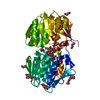
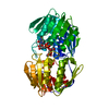
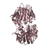
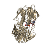
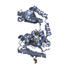
- PDB-2o0x: Mycobacterium tuberculosis epsp synthase in complex with intermediate -
+ Open data

- Basic information
| Entry | Database: PDB / ID: 2o0x | ||||||
|---|---|---|---|---|---|---|---|
| Title | Mycobacterium tuberculosis epsp synthase in complex with intermediate | ||||||
 Components Components | 3-phosphoshikimate 1-carboxyvinyltransferase | ||||||
 Keywords Keywords | TRANSFERASE / SHIKIMATE PATHWAY / EPSP SYNTHASE / M.TUBERCULOSIS / Structural Genomics / Mycobacterium Tuberculosis Structural Proteomics Project / XMTB | ||||||
| Function / homology |  Function and homology information Function and homology information3-phosphoshikimate 1-carboxyvinyltransferase / 3-phosphoshikimate 1-carboxyvinyltransferase activity / Chorismate via Shikimate Pathway / chorismate biosynthetic process / aromatic amino acid biosynthetic process / amino acid biosynthetic process / plasma membrane / cytoplasm / cytosol Similarity search - Function | ||||||
| Biological species |   Mycobacterium tuberculosis (bacteria) Mycobacterium tuberculosis (bacteria) | ||||||
| Method |  X-RAY DIFFRACTION / SYNCHROTRON / FOURIER SYNTHESIS / Resolution: 1.96 Å X-RAY DIFFRACTION / SYNCHROTRON / FOURIER SYNTHESIS / Resolution: 1.96 Å | ||||||
 Authors Authors | Kachalova, G.S. / Bartunik, H.D. / Mycobacterium Tuberculosis Structural Proteomics Project (XMTB) | ||||||
 Citation Citation | Journal: To be Published Title: Complexes of 3-PHOSPHOSHIKIMATE 1-CARBOXYVINYLTRANSFERASE Authors: Kachalova, G.S. / Burenkov, G.P. / Strizhov, N.I. / Brunning, M.G. / Bartunik, H.D. | ||||||
| History |
|
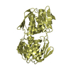
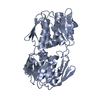
- Structure visualization
| Structure viewer | Molecule: MolmilJmol/JSmol |
|---|
- Downloads & links
-Download
| PDBx/mmCIF format | 2o0x.cif.gz | 114.5 KB | Display | PDBx/mmCIF format |
|---|---|---|---|---|
| PDB format | pdb2o0x.ent.gz | 85 KB | Display | PDB format |
| PDBx/mmJSON format | 2o0x.json.gz | Tree view | PDBx/mmJSON format | |
| Others |  Other downloads Other downloads |
-Validation report
| Arichive directory | https://data.pdbj.org/pub/pdb/validation_reports/o0/2o0xftp://data.pdbj.org/pub/pdb/validation_reports/o0/2o0x | HTTPS FTP |
|---|
-Related structure data
| Related structure data |  2o0dSC  2o0eC 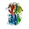 2o0zC  2o15C S: Starting model for refinement C: citing same article ( |
|---|---|
| Similar structure data | |
| Other databases |


-Links
 PDBj
PDBj

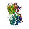
- Assembly
| Deposited unit | 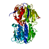
| ||||||||
|---|---|---|---|---|---|---|---|---|---|
| 1 |
| ||||||||
| Unit cell |
|
-Components
| #1: Protein | Mass: 46473.656 Da / Num. of mol.: 1 Source method: isolated from a genetically manipulated source Source: (gene. exp.) Mycobacterium tuberculosis (bacteria) / Gene: aroA / Plasmid: p7b, pET24b / Production host: References: UniProt: P22487, UniProt: P9WPY5*PLUS, 3-phosphoshikimate 1-carboxyvinyltransferase | ||||
|---|---|---|---|---|---|
| #2: Chemical |   Mass: 96.063 Da / Num. of mol.: 2 / Source method: obtained synthetically / Formula: SO4 Mass: 96.063 Da / Num. of mol.: 2 / Source method: obtained synthetically / Formula: SO4#3: Chemical | ChemComp-SKP / | 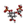  Mass: 422.173 Da / Num. of mol.: 1 / Source method: obtained synthetically / Formula: C10H16O14P2 Mass: 422.173 Da / Num. of mol.: 1 / Source method: obtained synthetically / Formula: C10H16O14P2#4: Water | ChemComp-HOH / |  Mass: 18.015 Da / Num. of mol.: 662 / Source method: isolated from a natural source / Formula: H2O Mass: 18.015 Da / Num. of mol.: 662 / Source method: isolated from a natural source / Formula: H2O |
-Experimental details
-Experiment
| Experiment | Method: X-RAY DIFFRACTION / Number of used crystals: 1 |
|---|
- Sample preparation
| Crystal | Density Matthews: 1.86 Å3/Da / Density % sol: 40.23 % |
|---|---|
| Crystal grow | Temperature: 293 K / Method: vapor diffusion, sitting drop / pH: 7.2 Details: 0.2M Ammonium sulfate ; 30%PEG 6000; 5mM S3P, pH 7.2, VAPOR DIFFUSION, SITTING DROP, temperature 293K |
-Data collection
| Diffraction | Mean temperature: 100 K |
|---|---|
| Diffraction source | Source: SYNCHROTRON / Site: MPG/DESY, HAMBURG  / Beamline: BW6 / Wavelength: 1.05 Å / Beamline: BW6 / Wavelength: 1.05 Å |
| Detector | Type: MAR CCD 165 mm / Detector: CCD / Date: Nov 25, 2004 / Details: Au-coated planar; toroidal mirrors |
| Radiation | Monochromator: Si111 / Protocol: SINGLE WAVELENGTH / Monochromatic (M) / Laue (L): M / Scattering type: x-ray |
| Radiation wavelength | Wavelength: 1.05 Å / Relative weight: 1 |
| Reflection | Resolution: 1.95→20 Å / Num. all: 28183 / Num. obs: 27055 / % possible obs: 95.9 % / Observed criterion σ(I): 2 / Redundancy: 5.5 % / Biso Wilson estimate: 24.1 Å2 / Rmerge(I) obs: 0.103 / Net I/σ(I): 13 |
| Reflection shell | Resolution: 1.95→1.98 Å / Redundancy: 5 % / Rmerge(I) obs: 0.65 / Mean I/σ(I) obs: 2 / Num. unique all: 1104 / % possible all: 89 |
- Processing
| Software |
| |||||||||||||||||||||||||||||||||
|---|---|---|---|---|---|---|---|---|---|---|---|---|---|---|---|---|---|---|---|---|---|---|---|---|---|---|---|---|---|---|---|---|---|---|
| Refinement | Method to determine structure: FOURIER SYNTHESIS Starting model: PDB ENTRY 2O0D Resolution: 1.96→10 Å / Num. parameters: 15858 / Num. restraintsaints: 14301 / Isotropic thermal model: isotropic / σ(F): 0 / Stereochemistry target values: ENGH AND HUBER
| |||||||||||||||||||||||||||||||||
| Displacement parameters | Biso mean: 23.9 Å2 | |||||||||||||||||||||||||||||||||
| Refine analyze | Num. disordered residues: 63 / Occupancy sum hydrogen: 0 / Occupancy sum non hydrogen: 3698.8 | |||||||||||||||||||||||||||||||||
| Refinement step | Cycle: LAST / Resolution: 1.96→10 Å
| |||||||||||||||||||||||||||||||||
| Refine LS restraints |
| |||||||||||||||||||||||||||||||||
| LS refinement shell | Resolution: 1.96→1.98 Å
|
