 Movie
Movie Controller


+ Open data

- Basic information
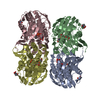
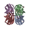
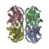
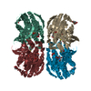
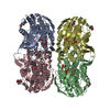
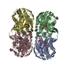
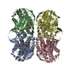
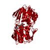
| Entry | Database: PDB / ID: 3v4t | ||||||
|---|---|---|---|---|---|---|---|
| Title | E. cloacae C115D MURA liganded with UNAG | ||||||
 Components Components | UDP-N-acetylglucosamine 1-carboxyvinyltransferase | ||||||
 Keywords Keywords | TRANSFERASE / MURA / CLOSE ENZYME STATE / CELL WALL / BIOGENESIS/DEGRADATION / PEPTIDOGLYCAN SYNTHESIS | ||||||
| Function / homology |  Function and homology information Function and homology informationUDP-N-acetylglucosamine 1-carboxyvinyltransferase activity / UDP-N-acetylgalactosamine biosynthetic process / UDP-N-acetylglucosamine 1-carboxyvinyltransferase / peptidoglycan biosynthetic process / cell wall organization / regulation of cell shape / cell division / cytoplasm Similarity search - Function | ||||||
| Biological species |  Enterobacter cloacae (bacteria) Enterobacter cloacae (bacteria) | ||||||
| Method |  X-RAY DIFFRACTION / MOLECULAR REPLACEMENT / Resolution: 2.5 Å X-RAY DIFFRACTION / MOLECULAR REPLACEMENT / Resolution: 2.5 Å | ||||||
 Authors Authors | Zhu, J.-Y. / Yang, Y. / Schonbrunn, E. | ||||||
 Citation Citation | Journal: J.Biol.Chem. / Year: 2012 Title: Functional Consequence of Covalent Reaction of Phosphoenolpyruvate with UDP-N-acetylglucosamine 1-Carboxyvinyltransferase (MurA). Authors: Zhu, J.Y. / Yang, Y. / Han, H. / Betzi, S. / Olesen, S.H. / Marsilio, F. / Schonbrunn, E. | ||||||
| History |
| ||||||
| Remark 999 | IAS67 FORMS AN ISOPEPTIDIC BOND AND IS A RESULT OF POSTTRANSLATIONAL MODIFICATION OF ASN67 |
- Structure visualization
| Structure viewer | Molecule: MolmilJmol/JSmol |
|---|
- Downloads & links
-Download
| PDBx/mmCIF format | 3v4t.cif.gz | 638 KB | Display | PDBx/mmCIF format |
|---|---|---|---|---|
| PDB format | pdb3v4t.ent.gz | 527.6 KB | Display | PDB format |
| PDBx/mmJSON format | 3v4t.json.gz | Tree view | PDBx/mmJSON format | |
| Others |  Other downloads Other downloads |
-Validation report
| Arichive directory | https://data.pdbj.org/pub/pdb/validation_reports/v4/3v4tftp://data.pdbj.org/pub/pdb/validation_reports/v4/3v4t | HTTPS FTP |
|---|
-Related structure data
| Related structure data |  3spbC 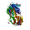 3su9SC  3swaC  3swdC 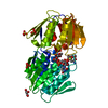 3sweC  3swgC 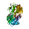 3swiC  3swqC  3upkC  3v5vC C: citing same article ( S: Starting model for refinement |
|---|---|
| Similar structure data |
-Links
 PDBj
PDBj
- Assembly
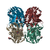
| Deposited unit | 
| ||||||||
|---|---|---|---|---|---|---|---|---|---|
| 1 | 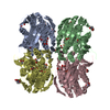
| ||||||||
| 2 | 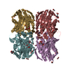
| ||||||||
| Unit cell |
|
-Components
| #1: Protein | Mass: 44841.352 Da / Num. of mol.: 8 / Mutation: C115D Source method: isolated from a genetically manipulated source Source: (gene. exp.) Enterobacter cloacae (bacteria) / Strain: ATCC 13047 / DSM 30054 / NBRC 13535 / NCDC 279-56 / Gene: ECL_04571, murA, murZ / Plasmid: PET9D / Production host: References: UniProt: P33038, UDP-N-acetylglucosamine 1-carboxyvinyltransferase #2: Chemical | ChemComp-UD1 /   Mass: 607.354 Da / Num. of mol.: 8 / Source method: obtained synthetically / Formula: C17H27N3O17P2 Mass: 607.354 Da / Num. of mol.: 8 / Source method: obtained synthetically / Formula: C17H27N3O17P2#3: Chemical | ChemComp-ACT /   Mass: 59.044 Da / Num. of mol.: 20 / Source method: obtained synthetically / Formula: C2H3O2 Mass: 59.044 Da / Num. of mol.: 20 / Source method: obtained synthetically / Formula: C2H3O2#4: Chemical | ChemComp-EDO /   Mass: 62.068 Da / Num. of mol.: 44 / Source method: obtained synthetically / Formula: C2H6O2 Mass: 62.068 Da / Num. of mol.: 44 / Source method: obtained synthetically / Formula: C2H6O2#5: Water | ChemComp-HOH / |  Mass: 18.015 Da / Num. of mol.: 470 / Source method: isolated from a natural source / Formula: H2O Mass: 18.015 Da / Num. of mol.: 470 / Source method: isolated from a natural source / Formula: H2OHas protein modification | Y | |
|---|
-Experimental details
-Experiment
| Experiment | Method: X-RAY DIFFRACTION / Number of used crystals: 1 |
|---|
- Sample preparation
| Crystal | Density Matthews: 2.28 Å3/Da / Density % sol: 46.06 % |
|---|---|
| Crystal grow | Temperature: 291 K / pH: 7.5 Details: 20 MG/ML MURA C115D, 2.5 MM UNAG, 25 MM HEPES PH 7.5, 50 MM BIS-TRIS PH 5.5, 100 MM AMMONIUM ACETATE, 12.5 % PEG 3350, VAPOR DIFFUSION, HANGING DROP, temperature 291K |
-Data collection
| Diffraction | Mean temperature: 93 K |
|---|---|
| Diffraction source | Source: ROTATING ANODE / Type: RIGAKU MICROMAX-007 HF / Wavelength: 1.54178 |
| Detector | Type: RIGAKU SATURN 944+ / Detector: CCD / Date: Feb 15, 2011 / Details: MIRRORS |
| Radiation | Monochromator: MIRRORS / Protocol: SINGLE WAVELENGTH / Monochromatic (M) / Laue (L): M / Scattering type: x-ray |
| Radiation wavelength | Wavelength: 1.54178 Å / Relative weight: 1 |
| Reflection | Resolution: 2.5→20 Å / Num. obs: 110457 / % possible obs: 99.5 % / Observed criterion σ(I): -3 / Redundancy: 3.7 % / Biso Wilson estimate: 37.9 Å2 / Rmerge(I) obs: 0.102 / Rsym value: 0.052 / Net I/σ(I): 19.7 |
| Reflection shell | Resolution: 2.5→2.6 Å / Redundancy: 3.7 % / Rmerge(I) obs: 0.425 / Mean I/σ(I) obs: 4.5 / Rsym value: 0.305 / % possible all: 99.7 |
- Processing
| Software |
| ||||||||||||||||||||||||||||||||||||||||||||||||||||||||||||||||||||||||||||||||
|---|---|---|---|---|---|---|---|---|---|---|---|---|---|---|---|---|---|---|---|---|---|---|---|---|---|---|---|---|---|---|---|---|---|---|---|---|---|---|---|---|---|---|---|---|---|---|---|---|---|---|---|---|---|---|---|---|---|---|---|---|---|---|---|---|---|---|---|---|---|---|---|---|---|---|---|---|---|---|---|---|---|
| Refinement | Method to determine structure: MOLECULAR REPLACEMENT Starting model: 3SU9 Resolution: 2.5→19.77 Å / Rfactor Rfree error: 0.008 / Data cutoff high absF: 3644161.9 / Data cutoff low absF: 0 / Isotropic thermal model: RESTRAINED / Cross valid method: THROUGHOUT / σ(F): 0 / Stereochemistry target values: ENGH & HUBER / Details: BULK SOLVENT MODEL USED
| ||||||||||||||||||||||||||||||||||||||||||||||||||||||||||||||||||||||||||||||||
| Solvent computation | Solvent model: FLAT MODEL / Bsol: 43.99 Å2 / ksol: 0.4 e/Å3 | ||||||||||||||||||||||||||||||||||||||||||||||||||||||||||||||||||||||||||||||||
| Displacement parameters | Biso mean: 42.4 Å2
| ||||||||||||||||||||||||||||||||||||||||||||||||||||||||||||||||||||||||||||||||
| Refine analyze |
| ||||||||||||||||||||||||||||||||||||||||||||||||||||||||||||||||||||||||||||||||
| Refinement step | Cycle: LAST / Resolution: 2.5→19.77 Å
| ||||||||||||||||||||||||||||||||||||||||||||||||||||||||||||||||||||||||||||||||
| Refine LS restraints |
| ||||||||||||||||||||||||||||||||||||||||||||||||||||||||||||||||||||||||||||||||
| Refine LS restraints NCS | NCS model details: NONE | ||||||||||||||||||||||||||||||||||||||||||||||||||||||||||||||||||||||||||||||||
| LS refinement shell | Resolution: 2.5→2.66 Å / Rfactor Rfree error: 0.023 / Total num. of bins used: 6
|
