 Movie
Movie Controller

[English] 日本語
 Yorodumi
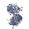
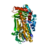
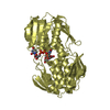
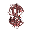
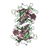
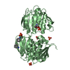
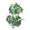
- PDB-3swg: AQUIFEX AEOLICUS MurA in complex with UDP-N-acetylmuramic acid an... -
+ Open data

- Basic information
| Entry | Database: PDB / ID: 3swg | ||||||
|---|---|---|---|---|---|---|---|
| Title | AQUIFEX AEOLICUS MurA in complex with UDP-N-acetylmuramic acid and covalent adduct of PEP with Cys124 | ||||||
 Components Components | UDP-N-acetylglucosamine 1-carboxyvinyltransferase | ||||||
 Keywords Keywords | TRANSFERASE / MURA / CLOSE ENZYME STATE / CELL WALL / BIOGENESIS/DEGRADATION / PEPTIDOGLYCAN SYNTHESIS | ||||||
| Function / homology |  Function and homology information Function and homology informationUDP-N-acetylglucosamine 1-carboxyvinyltransferase activity / UDP-N-acetylgalactosamine biosynthetic process / UDP-N-acetylglucosamine 1-carboxyvinyltransferase / peptidoglycan biosynthetic process / cell wall organization / regulation of cell shape / cell division / cytoplasm Similarity search - Function | ||||||
| Biological species |   Aquifex aeolicus (bacteria) Aquifex aeolicus (bacteria) | ||||||
| Method |  X-RAY DIFFRACTION / MOLECULAR REPLACEMENT / Resolution: 1.81 Å X-RAY DIFFRACTION / MOLECULAR REPLACEMENT / Resolution: 1.81 Å | ||||||
 Authors Authors | Zhu, J.-Y. / Schonbrunn, E. | ||||||
 Citation Citation | Journal: To be Published Title: Crystal structure of UDP-N-acetylglucosamine 1-carboxyvinyltransferase from Aquifex aeolicus VF5 Authors: Kitamura, Y. / Yokoyama, S. / Kuramitsu, S. | ||||||
| History |
| ||||||
| Remark 0 | THIS ENTRY 3SWG REFLECTS AN ALTERNATIVE MODELING OF THE ORIGINAL STRUCTURAL DATA R2YVWSF DETERMINED ...THIS ENTRY 3SWG REFLECTS AN ALTERNATIVE MODELING OF THE ORIGINAL STRUCTURAL DATA R2YVWSF DETERMINED BY AUTHORS OF THE PDB ENTRY 2YVW: AUTHOR Y.KITAMURA,S.YOKOYAMA,S.KURAMITSU | ||||||
| Remark 200 | AUTHOR USED THE SF DATA FROM ENTRY 2YVW. |
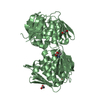
- Structure visualization
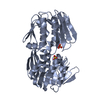
| Structure viewer | Molecule: MolmilJmol/JSmol |
|---|
- Downloads & links
-Download
| PDBx/mmCIF format | 3swg.cif.gz | 107.5 KB | Display | PDBx/mmCIF format |
|---|---|---|---|---|
| PDB format | pdb3swg.ent.gz | 81.4 KB | Display | PDB format |
| PDBx/mmJSON format | 3swg.json.gz | Tree view | PDBx/mmJSON format | |
| Others |  Other downloads Other downloads |
-Validation report
| Arichive directory | https://data.pdbj.org/pub/pdb/validation_reports/sw/3swgftp://data.pdbj.org/pub/pdb/validation_reports/sw/3swg | HTTPS FTP |
|---|
-Related structure data
| Related structure data |  3spbC 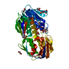 3su9C  3swaC  3swdC 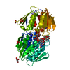 3sweC 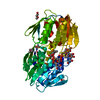 3swiC  3swqC  3upkC  3v4tC  3v5vC 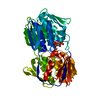 2yvwS S: Starting model for refinement C: citing same article ( |
|---|---|
| Similar structure data |

-Links
 PDBj
PDBj
- Assembly
| Deposited unit | 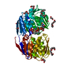
| ||||||||
|---|---|---|---|---|---|---|---|---|---|
| 1 |
| ||||||||
| Unit cell |
| ||||||||
| Components on special symmetry positions |
|
-Components
-Protein , 1 types, 1 molecules A
| #1: Protein | Mass: 47490.539 Da / Num. of mol.: 1 Source method: isolated from a genetically manipulated source Source: (gene. exp.) Aquifex aeolicus (bacteria) / Strain: VF5 / Gene: murA, aq_1281 / Plasmid: PET-21A / Production host: References: UniProt: O67315, UDP-N-acetylglucosamine 1-carboxyvinyltransferase |
|---|
-Non-polymers , 6 types, 366 molecules 


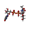

| #2: Chemical |  Mass: 194.226 Da / Num. of mol.: 3 / Source method: obtained synthetically / Formula: C8H18O5 / Comment: precipitant*YM Mass: 194.226 Da / Num. of mol.: 3 / Source method: obtained synthetically / Formula: C8H18O5 / Comment: precipitant*YM#3: Chemical | ChemComp-PGE / |  Mass: 150.173 Da / Num. of mol.: 1 / Source method: obtained synthetically / Formula: C6H14O4 Mass: 150.173 Da / Num. of mol.: 1 / Source method: obtained synthetically / Formula: C6H14O4#4: Chemical |  Mass: 106.120 Da / Num. of mol.: 2 / Source method: obtained synthetically / Formula: C4H10O3 Mass: 106.120 Da / Num. of mol.: 2 / Source method: obtained synthetically / Formula: C4H10O3#5: Chemical | ChemComp-EDO /  Mass: 62.068 Da / Num. of mol.: 11 / Source method: obtained synthetically / Formula: C2H6O2 Mass: 62.068 Da / Num. of mol.: 11 / Source method: obtained synthetically / Formula: C2H6O2#6: Chemical | ChemComp-EPZ / ( |  Mass: 679.416 Da / Num. of mol.: 1 / Source method: obtained synthetically / Formula: C20H31N3O19P2 Mass: 679.416 Da / Num. of mol.: 1 / Source method: obtained synthetically / Formula: C20H31N3O19P2#7: Water | ChemComp-HOH / | Mass: 18.015 Da / Num. of mol.: 348 / Source method: isolated from a natural source / Formula: H2O |
|---|
-Details
| Has protein modification | Y |
|---|
-Experimental details
-Experiment
| Experiment | Method: X-RAY DIFFRACTION |
|---|
- Sample preparation
| Crystal | Density Matthews: 3.08 Å3/Da / Density % sol: 60.06 % |
|---|
-Data collection
| Radiation | Scattering type: x-ray |
|---|---|
| Radiation wavelength | Relative weight: 1 |
- Processing
| Software | Name: CNS / Version: 1.3 / Classification: refinement | ||||||||||||||||||||||||||||||||||||||||||||||||||||||||||||||||||||||||||||||||
|---|---|---|---|---|---|---|---|---|---|---|---|---|---|---|---|---|---|---|---|---|---|---|---|---|---|---|---|---|---|---|---|---|---|---|---|---|---|---|---|---|---|---|---|---|---|---|---|---|---|---|---|---|---|---|---|---|---|---|---|---|---|---|---|---|---|---|---|---|---|---|---|---|---|---|---|---|---|---|---|---|---|
| Refinement | Method to determine structure: MOLECULAR REPLACEMENT Starting model: PDB entry 2YVW Resolution: 1.81→43.64 Å / Rfactor Rfree error: 0.004 / Data cutoff high absF: 91849.69 / Data cutoff low absF: 0 / Isotropic thermal model: RESTRAINED / Cross valid method: THROUGHOUT / σ(F): 0 / Details: BULK SOLVENT MODEL USED
| ||||||||||||||||||||||||||||||||||||||||||||||||||||||||||||||||||||||||||||||||
| Solvent computation | Solvent model: FLAT MODEL / Bsol: 60.7235 Å2 / ksol: 0.42 e/Å3 | ||||||||||||||||||||||||||||||||||||||||||||||||||||||||||||||||||||||||||||||||
| Displacement parameters | Biso mean: 24.1 Å2
| ||||||||||||||||||||||||||||||||||||||||||||||||||||||||||||||||||||||||||||||||
| Refine analyze |
| ||||||||||||||||||||||||||||||||||||||||||||||||||||||||||||||||||||||||||||||||
| Refinement step | Cycle: LAST / Resolution: 1.81→43.64 Å
| ||||||||||||||||||||||||||||||||||||||||||||||||||||||||||||||||||||||||||||||||
| Refine LS restraints |
| ||||||||||||||||||||||||||||||||||||||||||||||||||||||||||||||||||||||||||||||||
| LS refinement shell | Resolution: 1.81→1.91 Å / Rfactor Rfree error: 0.013 / Total num. of bins used: 6
| ||||||||||||||||||||||||||||||||||||||||||||||||||||||||||||||||||||||||||||||||
| Xplor file |
|
