 Movie
Movie Controller

[English] 日本語
 Yorodumi
- PDB-3e0q: Crystal structure of Schistosoma mansoni purine nucleoside phosph... -
+ Open data

- Basic information
| Entry | Database: PDB / ID: 3e0q | ||||||
|---|---|---|---|---|---|---|---|
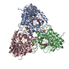
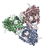
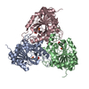
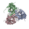
| Title | Crystal structure of Schistosoma mansoni purine nucleoside phosphorylase complexed with a novel monocyclic inhibitor | ||||||
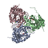
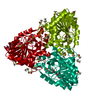
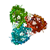
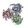
 Components Components | Purine-nucleoside phosphorylase | ||||||
 Keywords Keywords | TRANSFERASE / Monocyclic inhibitor / Glycosyltransferase | ||||||
| Function / homology |  Function and homology information Function and homology informationnucleoside metabolic process / guanosine phosphorylase activity / purine-nucleoside phosphorylase / purine-nucleoside phosphorylase activity / cytoplasm Similarity search - Function | ||||||
| Biological species |  | ||||||
| Method |  X-RAY DIFFRACTION / SYNCHROTRON / FOURIER SYNTHESIS / molecular replacement / Resolution: 1.9 Å X-RAY DIFFRACTION / SYNCHROTRON / FOURIER SYNTHESIS / molecular replacement / Resolution: 1.9 Å | ||||||
 Authors Authors | Pereira, H.M. / Berdini, V. / Ferri, M.R. / Cleasby, A. / Garratt, R.C. | ||||||
 Citation Citation | Journal: Acta Trop. / Year: 2010 Title: Crystal structure of Schistosoma purine nucleoside phosphorylase complexed with a novel monocyclic inhibitor. Authors: Pereira, H.M. / Berdini, V. / Ferri, M.R. / Cleasby, A. / Garratt, R.C. #1: Journal: J.Mol.Biol. / Year: 2005Title: Structures for the potential drug target purine nucleoside phosphorylase from Schistosoma mansoni causal agent of schistosomiasis. Authors: Pereira, H.D. / Franco, G.R. / Cleasby, A. / Garratt, R.C. | ||||||
| History |
|
- Structure visualization
| Structure viewer | Molecule: MolmilJmol/JSmol |
|---|
- Downloads & links
-Download
| PDBx/mmCIF format | 3e0q.cif.gz | 180.3 KB | Display | PDBx/mmCIF format |
|---|---|---|---|---|
| PDB format | pdb3e0q.ent.gz | 143.3 KB | Display | PDB format |
| PDBx/mmJSON format | 3e0q.json.gz | Tree view | PDBx/mmJSON format | |
| Others |  Other downloads Other downloads |
-Validation report
| Arichive directory | https://data.pdbj.org/pub/pdb/validation_reports/e0/3e0qftp://data.pdbj.org/pub/pdb/validation_reports/e0/3e0q | HTTPS FTP |
|---|
-Related structure data
| Related structure data |  1tcvS S: Starting model for refinement |
|---|---|
| Similar structure data |


-Links
 PDBj
PDBj- Assembly
| Deposited unit | 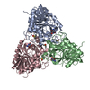
| ||||||||
|---|---|---|---|---|---|---|---|---|---|
| 1 |
| ||||||||
| Unit cell |
|
-Components
| #1: Protein | Mass: 31197.254 Da / Num. of mol.: 3 Source method: isolated from a genetically manipulated source Source: (gene. exp.)  References: UniProt: Q9BMI9, purine-nucleoside phosphorylase #2: Chemical | ChemComp-DMS /   Mass: 78.133 Da / Num. of mol.: 4 / Source method: obtained synthetically / Formula: C2H6OS / Comment: DMSO, precipitant*YM Mass: 78.133 Da / Num. of mol.: 4 / Source method: obtained synthetically / Formula: C2H6OS / Comment: DMSO, precipitant*YM#3: Chemical | 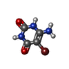  Mass: 205.997 Da / Num. of mol.: 3 / Source method: obtained synthetically / Formula: C4H4BrN3O2 Mass: 205.997 Da / Num. of mol.: 3 / Source method: obtained synthetically / Formula: C4H4BrN3O2#4: Water | ChemComp-HOH / |  Mass: 18.015 Da / Num. of mol.: 584 / Source method: isolated from a natural source / Formula: H2O Mass: 18.015 Da / Num. of mol.: 584 / Source method: isolated from a natural source / Formula: H2O |
|---|
-Experimental details
-Experiment
| Experiment | Method: X-RAY DIFFRACTION / Number of used crystals: 1 |
|---|
- Sample preparation
| Crystal | Density Matthews: 2.02 Å3/Da / Density % sol: 38.98 % |
|---|---|
| Crystal grow | Temperature: 277 K / Method: vapor diffusion, hanging drop Details: 18-20% PEG 1500, 20% Glycerol, 32mM Sodium acetate, pH 4.9-5.0, VAPOR DIFFUSION, HANGING DROP, temperature 277K |
-Data collection
| Diffraction | Mean temperature: 100 K | ||||||||||||||||||||||||||||||||||||||||||||||||||||||||||||||||||||||||||||||||||||||||
|---|---|---|---|---|---|---|---|---|---|---|---|---|---|---|---|---|---|---|---|---|---|---|---|---|---|---|---|---|---|---|---|---|---|---|---|---|---|---|---|---|---|---|---|---|---|---|---|---|---|---|---|---|---|---|---|---|---|---|---|---|---|---|---|---|---|---|---|---|---|---|---|---|---|---|---|---|---|---|---|---|---|---|---|---|---|---|---|---|---|
| Diffraction source | Source: SYNCHROTRON / Site: SRS  / Beamline: PX14.2 / Wavelength: 0.98 Å / Beamline: PX14.2 / Wavelength: 0.98 Å | ||||||||||||||||||||||||||||||||||||||||||||||||||||||||||||||||||||||||||||||||||||||||
| Detector | Type: ADSC QUANTUM 210 / Detector: CCD / Date: Mar 27, 2002 | ||||||||||||||||||||||||||||||||||||||||||||||||||||||||||||||||||||||||||||||||||||||||
| Radiation | Protocol: SINGLE WAVELENGTH / Monochromatic (M) / Laue (L): M / Scattering type: x-ray | ||||||||||||||||||||||||||||||||||||||||||||||||||||||||||||||||||||||||||||||||||||||||
| Radiation wavelength | Wavelength: 0.98 Å / Relative weight: 1 | ||||||||||||||||||||||||||||||||||||||||||||||||||||||||||||||||||||||||||||||||||||||||
| Reflection | Resolution: 1.9→44.46 Å / Num. obs: 56943 / % possible obs: 94.8 % / Redundancy: 2.6 % / Rmerge(I) obs: 0.071 / Rsym value: 0.071 | ||||||||||||||||||||||||||||||||||||||||||||||||||||||||||||||||||||||||||||||||||||||||
| Reflection shell |
|
-Phasing
| Phasing | Method: molecular replacement |
|---|
- Processing
| Software |
| |||||||||||||||||||||||||||||||||||||||||||||||||||||||||||||||||||||||||||||||||||||||||||||||||||||||||||||||||||||||||||||||||||||||||||||||||||
|---|---|---|---|---|---|---|---|---|---|---|---|---|---|---|---|---|---|---|---|---|---|---|---|---|---|---|---|---|---|---|---|---|---|---|---|---|---|---|---|---|---|---|---|---|---|---|---|---|---|---|---|---|---|---|---|---|---|---|---|---|---|---|---|---|---|---|---|---|---|---|---|---|---|---|---|---|---|---|---|---|---|---|---|---|---|---|---|---|---|---|---|---|---|---|---|---|---|---|---|---|---|---|---|---|---|---|---|---|---|---|---|---|---|---|---|---|---|---|---|---|---|---|---|---|---|---|---|---|---|---|---|---|---|---|---|---|---|---|---|---|---|---|---|---|---|---|---|---|
| Refinement | Method to determine structure: FOURIER SYNTHESIS Starting model: PDB entry 1TCV Resolution: 1.9→44.32 Å / Num. restraintsaints: 87047 / Occupancy max: 1 / Occupancy min: 0 / SU ML: 0.27 / σ(F): 1.34 / Stereochemistry target values: MAXIMUM LIKELIHOOD
| |||||||||||||||||||||||||||||||||||||||||||||||||||||||||||||||||||||||||||||||||||||||||||||||||||||||||||||||||||||||||||||||||||||||||||||||||||
| Solvent computation | Shrinkage radii: 0.9 Å / VDW probe radii: 1.11 Å / Solvent model: FLAT BULK SOLVENT MODEL / Bsol: 48.161 Å2 / ksol: 0.367 e/Å3 | |||||||||||||||||||||||||||||||||||||||||||||||||||||||||||||||||||||||||||||||||||||||||||||||||||||||||||||||||||||||||||||||||||||||||||||||||||
| Displacement parameters | Biso max: 60.82 Å2 / Biso mean: 20.249 Å2 / Biso min: 5.34 Å2
| |||||||||||||||||||||||||||||||||||||||||||||||||||||||||||||||||||||||||||||||||||||||||||||||||||||||||||||||||||||||||||||||||||||||||||||||||||
| Refinement step | Cycle: LAST / Resolution: 1.9→44.32 Å
| |||||||||||||||||||||||||||||||||||||||||||||||||||||||||||||||||||||||||||||||||||||||||||||||||||||||||||||||||||||||||||||||||||||||||||||||||||
| Refine LS restraints |
| |||||||||||||||||||||||||||||||||||||||||||||||||||||||||||||||||||||||||||||||||||||||||||||||||||||||||||||||||||||||||||||||||||||||||||||||||||
| LS refinement shell | Refine-ID: X-RAY DIFFRACTION / Total num. of bins used: 20
|
