 Movie
Movie Controller


+ Open data

- Basic information
| Entry | Database: PDB / ID: 3d4f | ||||||
|---|---|---|---|---|---|---|---|
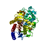
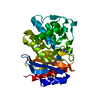
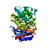
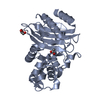
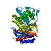
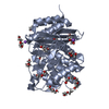
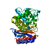
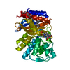
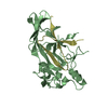
| Title | SHV-1 beta-lactamase complex with LN1-255 | ||||||
 Components Components | Beta-lactamase SHV-1 | ||||||
 Keywords Keywords | HYDROLASE / BETA-LACTAMASE INHIBITOR / DRUG DESIGN / Antibiotic resistance / Plasmid | ||||||
| Function / homology |  Function and homology information Function and homology informationbeta-lactam antibiotic catabolic process / beta-lactamase activity / beta-lactamase / response to antibiotic Similarity search - Function | ||||||
| Biological species |  Klebsiella pneumoniae (bacteria) Klebsiella pneumoniae (bacteria) | ||||||
| Method |  X-RAY DIFFRACTION / SYNCHROTRON / MOLECULAR REPLACEMENT / Resolution: 1.55 Å X-RAY DIFFRACTION / SYNCHROTRON / MOLECULAR REPLACEMENT / Resolution: 1.55 Å | ||||||
 Authors Authors | Pattanaik, P. / Bethel, C.R. / Hujer, A.M. / Bonomo, R.A. / Buynak, J.D. / van den Akker, F. | ||||||
 Citation Citation | Journal: J.Biol.Chem. / Year: 2009 Title: Strategic Design of an Effective {beta}-Lactamase Inhibitor: LN-1-255, A 6-ALKYLIDENE-2'-SUBSTITUTED PENICILLIN SULFONE Authors: Pattanaik, P. / Bethel, C.R. / Hujer, A.M. / Hujer, K.M. / Distler, A.M. / Taracila, M. / Anderson, V.E. / Fritsche, T.R. / Jones, R.N. / Pagadala, S.R. / van den Akker, F. / Buynak, J.D. / Bonomo, R.A. | ||||||
| History |
|
- Structure visualization
| Structure viewer | Molecule: MolmilJmol/JSmol |
|---|
- Downloads & links
-Download
| PDBx/mmCIF format | 3d4f.cif.gz | 73.9 KB | Display | PDBx/mmCIF format |
|---|---|---|---|---|
| PDB format | pdb3d4f.ent.gz | 53.3 KB | Display | PDB format |
| PDBx/mmJSON format | 3d4f.json.gz | Tree view | PDBx/mmJSON format | |
| Others |  Other downloads Other downloads |
-Validation report
| Arichive directory | https://data.pdbj.org/pub/pdb/validation_reports/d4/3d4fftp://data.pdbj.org/pub/pdb/validation_reports/d4/3d4f | HTTPS FTP |
|---|
-Related structure data
| Related structure data | |
|---|---|
| Similar structure data |


-Links
 PDBj
PDBj


- Assembly
| Deposited unit | 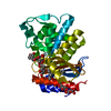
| ||||||||
|---|---|---|---|---|---|---|---|---|---|
| 1 |
| ||||||||
| Unit cell |
| ||||||||
| Details | Monomer |
-Components
| #1: Protein | Mass: 31259.988 Da / Num. of mol.: 1 Source method: isolated from a genetically manipulated source Source: (gene. exp.) Klebsiella pneumoniae (bacteria) / Gene: bla, shv1 / Plasmid: PBC SK(-) / Production host: | ||||||
|---|---|---|---|---|---|---|---|
| #2: Chemical |   Mass: 508.600 Da / Num. of mol.: 2 / Source method: obtained synthetically / Formula: C24H44O11 Mass: 508.600 Da / Num. of mol.: 2 / Source method: obtained synthetically / Formula: C24H44O11#3: Chemical | ChemComp-LN1 / ( |   Mass: 490.483 Da / Num. of mol.: 1 / Source method: obtained synthetically / Formula: C22H22N2O9S Mass: 490.483 Da / Num. of mol.: 1 / Source method: obtained synthetically / Formula: C22H22N2O9S#4: Water | ChemComp-HOH / |  Mass: 18.015 Da / Num. of mol.: 248 / Source method: isolated from a natural source / Formula: H2O Mass: 18.015 Da / Num. of mol.: 248 / Source method: isolated from a natural source / Formula: H2OHas protein modification | Y | |
-Experimental details
-Experiment
| Experiment | Method: X-RAY DIFFRACTION / Number of used crystals: 1 |
|---|
- Sample preparation
| Crystal | Density Matthews: 1.84 Å3/Da / Density % sol: 33.11 % |
|---|---|
| Crystal grow | Temperature: 298 K / Method: vapor diffusion, sitting drop / pH: 7 Details: 30% PEG6000, 0.1M HEPES pH7. 0.56mM Cymal-6, VAPOR DIFFUSION, SITTING DROP, temperature 298K |
-Data collection
| Diffraction | Mean temperature: 100 K |
|---|---|
| Diffraction source | Source: SYNCHROTRON / Site: NSLS  / Beamline: X29A / Wavelength: 1.1 Å / Beamline: X29A / Wavelength: 1.1 Å |
| Detector | Type: ADSC QUANTUM 315 / Detector: CCD / Date: Mar 25, 2006 |
| Radiation | Monochromator: vertically focusing mirror and a horizontally focusing monochromator Protocol: SINGLE WAVELENGTH / Monochromatic (M) / Laue (L): M / Scattering type: x-ray |
| Radiation wavelength | Wavelength: 1.1 Å / Relative weight: 1 |
| Reflection | Resolution: 1.55→50 Å / Num. all: 34280 / Num. obs: 33525 / % possible obs: 97.8 % / Observed criterion σ(F): 0 / Observed criterion σ(I): -3 / Biso Wilson estimate: 16.8 Å2 / Rsym value: 0.101 / Net I/σ(I): 20.8 |
| Reflection shell | Resolution: 1.55→1.61 Å / Rmerge(I) obs: 0.41 / Mean I/σ(I) obs: 2.1 / % possible all: 96.3 |
- Processing
| Software |
| |||||||||||||||||||||||||
|---|---|---|---|---|---|---|---|---|---|---|---|---|---|---|---|---|---|---|---|---|---|---|---|---|---|---|
| Refinement | Method to determine structure: MOLECULAR REPLACEMENT Starting model: SHV-1 wt Resolution: 1.55→31.95 Å / Rfactor Rfree error: 0.005 / Data cutoff high absF: 1336274.31 / Data cutoff low absF: 0 / Isotropic thermal model: RESTRAINED / Cross valid method: THROUGHOUT / σ(F): 0 / Stereochemistry target values: Engh & Huber / Details: BULK SOLVENT MODEL USED
| |||||||||||||||||||||||||
| Solvent computation | Solvent model: FLAT MODEL / Bsol: 53.5903 Å2 / ksol: 0.35 e/Å3 | |||||||||||||||||||||||||
| Displacement parameters | Biso mean: 17 Å2
| |||||||||||||||||||||||||
| Refine analyze |
| |||||||||||||||||||||||||
| Refinement step | Cycle: LAST / Resolution: 1.55→31.95 Å
| |||||||||||||||||||||||||
| Refine LS restraints |
| |||||||||||||||||||||||||
| LS refinement shell | Resolution: 1.55→1.65 Å / Rfactor Rfree error: 0.017 / Total num. of bins used: 6
| |||||||||||||||||||||||||
| Xplor file |
|
