 Movie
Movie Controller

[English] 日本語
 Yorodumi
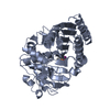
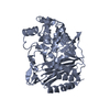
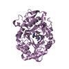
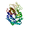
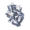
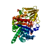
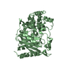
- PDB-1ga0: STRUCTURE OF THE E. CLOACAE GC1 BETA-LACTAMASE WITH A CEPHALOSPOR... -
+ Open data

- Basic information
| Entry | Database: PDB / ID: 1ga0 | ||||||
|---|---|---|---|---|---|---|---|
| Title | STRUCTURE OF THE E. CLOACAE GC1 BETA-LACTAMASE WITH A CEPHALOSPORIN SULFONE INHIBITOR | ||||||
 Components Components | BETA-LACTAMASE | ||||||
 Keywords Keywords | HYDROLASE / mixed alpha/beta / cephalosporinase / inhibition / conformational change / class C beta-lactamase | ||||||
| Function / homology |  Function and homology information Function and homology informationantibiotic catabolic process / beta-lactamase activity / beta-lactamase / outer membrane-bounded periplasmic space / response to antibiotic Similarity search - Function | ||||||
| Biological species |  Enterobacter cloacae (bacteria) Enterobacter cloacae (bacteria) | ||||||
| Method |  X-RAY DIFFRACTION / SYNCHROTRON / MOLECULAR REPLACEMENT / Resolution: 1.6 Å X-RAY DIFFRACTION / SYNCHROTRON / MOLECULAR REPLACEMENT / Resolution: 1.6 Å | ||||||
 Authors Authors | Crichlow, G.V. / Nukaga, M. / Buynak, J.D. / Knox, J.R. | ||||||
 Citation Citation | Journal: Biochemistry / Year: 2001 Title: Inhibition of class C beta-lactamases: structure of a reaction intermediate with a cephem sulfone. Authors: Crichlow, G.V. / Nukaga, M. / Doppalapudi, V.R. / Buynak, J.D. / Knox, J.R. | ||||||
| History |
|
- Structure visualization
| Structure viewer | Molecule: MolmilJmol/JSmol |
|---|
- Downloads & links
-Download
| PDBx/mmCIF format | 1ga0.cif.gz | 89.8 KB | Display | PDBx/mmCIF format |
|---|---|---|---|---|
| PDB format | pdb1ga0.ent.gz | 67.1 KB | Display | PDB format |
| PDBx/mmJSON format | 1ga0.json.gz | Tree view | PDBx/mmJSON format | |
| Others |  Other downloads Other downloads |
-Validation report
| Arichive directory | https://data.pdbj.org/pub/pdb/validation_reports/ga/1ga0ftp://data.pdbj.org/pub/pdb/validation_reports/ga/1ga0 | HTTPS FTP |
|---|
-Related structure data
| Related structure data | 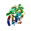 1gceS S: Starting model for refinement |
|---|---|
| Similar structure data |


-Links
 PDBj
PDBj


- Assembly
| Deposited unit | 
| ||||||||
|---|---|---|---|---|---|---|---|---|---|
| 1 |
| ||||||||
| Unit cell |
| ||||||||
| Components on special symmetry positions |
|
-Components
| #1: Protein | Mass: 39609.172 Da / Num. of mol.: 1 Source method: isolated from a genetically manipulated source Source: (gene. exp.) Enterobacter cloacae (bacteria) / Gene: BLAC / Plasmid: PCS101 / Production host: |
|---|---|
| #2: Chemical | ChemComp-NA /   Mass: 22.990 Da / Num. of mol.: 1 / Fragment: DVR-II-41S / Source method: obtained synthetically / Formula: Na Mass: 22.990 Da / Num. of mol.: 1 / Fragment: DVR-II-41S / Source method: obtained synthetically / Formula: Na |
| #3: Chemical | ChemComp-DVR /   Mass: 377.372 Da / Num. of mol.: 1 / Source method: obtained synthetically / Formula: C16H15N3O6S Mass: 377.372 Da / Num. of mol.: 1 / Source method: obtained synthetically / Formula: C16H15N3O6S |
| #4: Chemical | ChemComp-GOL /   Mass: 92.094 Da / Num. of mol.: 1 / Source method: obtained synthetically / Formula: C3H8O3 Mass: 92.094 Da / Num. of mol.: 1 / Source method: obtained synthetically / Formula: C3H8O3 |
| #5: Water | ChemComp-HOH /  Mass: 18.015 Da / Num. of mol.: 315 / Source method: isolated from a natural source / Formula: H2O Mass: 18.015 Da / Num. of mol.: 315 / Source method: isolated from a natural source / Formula: H2O |
| Has protein modification | Y |
-Experimental details
-Experiment
| Experiment | Method: X-RAY DIFFRACTION / Number of used crystals: 1 |
|---|
- Sample preparation
| Crystal | Density Matthews: 2.09 Å3/Da / Density % sol: 41.27 % | ||||||||||||||||||||
|---|---|---|---|---|---|---|---|---|---|---|---|---|---|---|---|---|---|---|---|---|---|
| Crystal grow | Temperature: 293 K / Method: vapor diffusion, sitting drop / pH: 7 Details: 6.3 mg/ml enzyme, 5.5% PEG 3350, 9 mM imidazole; over a reservoir of 24% PEG 3350. Crystal was reacted with 9 mM DVR-II-41s in 15 mM imidazole/28% PEG 3350, pH 7.0 for 3 hr. and then soaked ...Details: 6.3 mg/ml enzyme, 5.5% PEG 3350, 9 mM imidazole; over a reservoir of 24% PEG 3350. Crystal was reacted with 9 mM DVR-II-41s in 15 mM imidazole/28% PEG 3350, pH 7.0 for 3 hr. and then soaked in an identical solution for 35 min. The crystal was soaked in a cryo-protectant solution of 25% glycerol/20 mM HEPES (pH 7)/24% PEG for 2 min. prior to cryo-freezing., VAPOR DIFFUSION, SITTING DROP, temperature 293K | ||||||||||||||||||||
| Crystal grow | *PLUS | ||||||||||||||||||||
| Components of the solutions | *PLUS
|
-Data collection
| Diffraction | Mean temperature: 100 K |
|---|---|
| Diffraction source | Source: SYNCHROTRON / Site: CHESS  / Beamline: A1 / Wavelength: 0.935 Å / Beamline: A1 / Wavelength: 0.935 Å |
| Detector | Type: ADSC QUANTUM 4 / Detector: CCD / Date: Feb 10, 1999 |
| Radiation | Protocol: SINGLE WAVELENGTH / Monochromatic (M) / Laue (L): M / Scattering type: x-ray |
| Radiation wavelength | Wavelength: 0.935 Å / Relative weight: 1 |
| Reflection | Resolution: 1.6→50 Å / Num. obs: 29788 / % possible obs: 66.1 % / Observed criterion σ(I): -3 / Redundancy: 3.2 % / Biso Wilson estimate: 10.8 Å2 / Rmerge(I) obs: 0.087 / Net I/σ(I): 7.8 |
| Reflection shell | Resolution: 1.6→2.02 Å / Redundancy: 1.7 % / Rmerge(I) obs: 0.31 / Mean I/σ(I) obs: 2.7 / Num. unique all: 7159 / % possible all: 32.2 |
| Reflection | *PLUS Lowest resolution: 50 Å / Num. measured all: 94739 |
| Reflection shell | *PLUS % possible obs: 32.2 % / Num. unique obs: 7159 / Num. measured obs: 12241 |
- Processing
| Software |
| |||||||||||||||||||||||||
|---|---|---|---|---|---|---|---|---|---|---|---|---|---|---|---|---|---|---|---|---|---|---|---|---|---|---|
| Refinement | Method to determine structure: MOLECULAR REPLACEMENT Starting model: PDB entry 1GCE Resolution: 1.6→100 Å / Isotropic thermal model: restrained, isotropic / Cross valid method: THROUGHOUT / σ(F): 0 / σ(I): 0 Stereochemistry target values: Engh & Huber (for the protein) Details: Intermediate assigned occupancy of 1; however, it may be less. There are six alternate side chain conformations in the protein and one alternate conformation for atom C2 and the sulfinate ...Details: Intermediate assigned occupancy of 1; however, it may be less. There are six alternate side chain conformations in the protein and one alternate conformation for atom C2 and the sulfinate atoms of the inhibitor. A bulk solvent correction was used.
| |||||||||||||||||||||||||
| Displacement parameters | Biso mean: 14.7 Å2 | |||||||||||||||||||||||||
| Refine analyze |
| |||||||||||||||||||||||||
| Refinement step | Cycle: LAST / Resolution: 1.6→100 Å
| |||||||||||||||||||||||||
| Refine LS restraints |
| |||||||||||||||||||||||||
| Software | *PLUS Name: CNS / Version: 1 / Classification: refinement | |||||||||||||||||||||||||
| Refinement | *PLUS Highest resolution: 1.6 Å / Lowest resolution: 100 Å / σ(F): 0 | |||||||||||||||||||||||||
| Solvent computation | *PLUS | |||||||||||||||||||||||||
| Displacement parameters | *PLUS Biso mean: 14.7 Å2 | |||||||||||||||||||||||||
| Refine LS restraints | *PLUS Type: c_plane_restr / Dev ideal: 1 |
