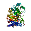
 Movie
Movie Controller


+ Open data

- Basic information
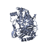
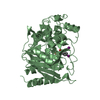
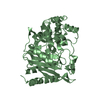
| Entry | Database: PDB / ID: 1gce | ||||||
|---|---|---|---|---|---|---|---|
| Title | STRUCTURE OF THE BETA-LACTAMASE OF ENTEROBACTER CLOACAE GC1 | ||||||
 Components Components | BETA-LACTAMASE | ||||||
 Keywords Keywords | HYDROLASE / BETA-LACTAM HYDROLASE / CEPHALOSPORINASE / DRUG DESIGN / EXTENDED-SPECTRUM BETA- LACTAMASE | ||||||
| Function / homology |  Function and homology information Function and homology informationantibiotic catabolic process / beta-lactamase activity / beta-lactamase / outer membrane-bounded periplasmic space / response to antibiotic Similarity search - Function | ||||||
| Biological species |  Enterobacter cloacae (bacteria) Enterobacter cloacae (bacteria) | ||||||
| Method |  X-RAY DIFFRACTION / SYNCHROTRON / MOLECULAR REPLACEMENT / Resolution: 1.8 Å X-RAY DIFFRACTION / SYNCHROTRON / MOLECULAR REPLACEMENT / Resolution: 1.8 Å | ||||||
 Authors Authors | Crichlow, G.V. / Kuzin, A.P. / Nukaga, M. / Sawai, T. / Knox, J.R. | ||||||
 Citation Citation | Journal: Biochemistry / Year: 1999 Title: Structure of the extended-spectrum class C beta-lactamase of Enterobacter cloacae GC1, a natural mutant with a tandem tripeptide insertion. Authors: Crichlow, G.V. / Kuzin, A.P. / Nukaga, M. / Mayama, K. / Sawai, T. / Knox, J.R. | ||||||
| History |
|
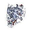
- Structure visualization
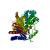
| Structure viewer | Molecule: MolmilJmol/JSmol |
|---|
- Downloads & links
-Download
| PDBx/mmCIF format | 1gce.cif.gz | 86.4 KB | Display | PDBx/mmCIF format |
|---|---|---|---|---|
| PDB format | pdb1gce.ent.gz | 64.2 KB | Display | PDB format |
| PDBx/mmJSON format | 1gce.json.gz | Tree view | PDBx/mmJSON format | |
| Others |  Other downloads Other downloads |
-Validation report
| Arichive directory | https://data.pdbj.org/pub/pdb/validation_reports/gc/1gceftp://data.pdbj.org/pub/pdb/validation_reports/gc/1gce | HTTPS FTP |
|---|
-Related structure data
| Related structure data |  2blt S: Starting model for refinement |
|---|---|
| Similar structure data |


-Links
 PDBj
PDBj
- Assembly
| Deposited unit | 
| ||||||||||||
|---|---|---|---|---|---|---|---|---|---|---|---|---|---|
| 1 |
| ||||||||||||
| Unit cell |
| ||||||||||||
| Components on special symmetry positions |
|
-Components
| #1: Protein | Mass: 39609.172 Da / Num. of mol.: 1 Source method: isolated from a genetically manipulated source Source: (gene. exp.) Enterobacter cloacae (bacteria) / Strain: GC1 / Cellular location: PERIPLASM / Plasmid: PTTQ18K-GC1 / Gene (production host): BLAC / Production host: |
|---|---|
| #2: Water | ChemComp-HOH /  Mass: 18.015 Da / Num. of mol.: 235 / Source method: isolated from a natural source / Formula: H2O Mass: 18.015 Da / Num. of mol.: 235 / Source method: isolated from a natural source / Formula: H2O |
-Experimental details
-Experiment
| Experiment | Method: X-RAY DIFFRACTION / Number of used crystals: 1 |
|---|
- Sample preparation
| Crystal | Density Matthews: 2.16 Å3/Da / Density % sol: 43 % Description: PH GIVEN IS THAT OF A SOLUTION SIMILAR TO THE RESERVOIR SOLUION. THE PH FALLS IN THE RANGE OF 4 - 6. | ||||||||||||||||||||||||
|---|---|---|---|---|---|---|---|---|---|---|---|---|---|---|---|---|---|---|---|---|---|---|---|---|---|
| Crystal grow | Temperature: 298 K / Method: vapor diffusion / pH: 5 Details: VAPOR DIFFUSION, WITH 6.7 OR 8.0 MG/ML PROTEIN IN 5-7.5% PEG8000, 20-25MM POTASSIUM PHOSPHATE (MONOBASIC), OVER 12.5 OR 15% PEG8000, 50MM POTASSIUM PHOSPHATE (MONOBASIC) RESERVOIR AT ROOM ...Details: VAPOR DIFFUSION, WITH 6.7 OR 8.0 MG/ML PROTEIN IN 5-7.5% PEG8000, 20-25MM POTASSIUM PHOSPHATE (MONOBASIC), OVER 12.5 OR 15% PEG8000, 50MM POTASSIUM PHOSPHATE (MONOBASIC) RESERVOIR AT ROOM TEMPERATURE., temperature 298K | ||||||||||||||||||||||||
| Crystal grow | *PLUS Method: vapor diffusion, sitting drop | ||||||||||||||||||||||||
| Components of the solutions | *PLUS
|
-Data collection
| Diffraction | Mean temperature: 100 K |
|---|---|
| Diffraction source | Source: SYNCHROTRON / Site: CHESS  / Beamline: A1 / Wavelength: 0.908 / Beamline: A1 / Wavelength: 0.908 |
| Detector | Type: ADSC / Detector: CCD / Date: Aug 1, 1997 |
| Radiation | Protocol: SINGLE WAVELENGTH / Monochromatic (M) / Laue (L): M / Scattering type: x-ray |
| Radiation wavelength | Wavelength: 0.908 Å / Relative weight: 1 |
| Reflection | Resolution: 1.8→99 Å / Num. obs: 31187 / % possible obs: 95.6 % / Observed criterion σ(I): -3 / Redundancy: 3.9 % / Rmerge(I) obs: 0.076 / Rsym value: 0.076 / Net I/σ(I): 11.1 |
| Reflection shell | Resolution: 1.8→1.86 Å / Redundancy: 1.79 % / Rmerge(I) obs: 0.142 / Mean I/σ(I) obs: 5 / Rsym value: 0.142 / % possible all: 80 |
| Reflection | *PLUS % possible obs: 96 % / Num. measured all: 122470 |
| Reflection shell | *PLUS % possible obs: 80 % / Num. unique obs: 2568 / Num. measured obs: 4591 / Mean I/σ(I) obs: 5.1 |
- Processing
| Software |
| ||||||||||||||||||||||||||||||||||||||||||||||||||||||||||||
|---|---|---|---|---|---|---|---|---|---|---|---|---|---|---|---|---|---|---|---|---|---|---|---|---|---|---|---|---|---|---|---|---|---|---|---|---|---|---|---|---|---|---|---|---|---|---|---|---|---|---|---|---|---|---|---|---|---|---|---|---|---|
| Refinement | Method to determine structure: MOLECULAR REPLACEMENT Starting model: 2BLT 2blt Resolution: 1.8→8 Å / Data cutoff high absF: 1000000 / Data cutoff low absF: 0 / Cross valid method: THROUGHOUT / σ(F): 0 Details: AFTER FINAL REFINEMENT, ONE WATER MOLECULE WAS DELETED FROM MODEL DUE TO B- FACTOR MORE THAN 55. AVG. B GIVEN REFLECTS STRUCTURE INCLUDING PROTEIN AND ONLY 235 WATER MOLECULES. THE FINAL ...Details: AFTER FINAL REFINEMENT, ONE WATER MOLECULE WAS DELETED FROM MODEL DUE TO B- FACTOR MORE THAN 55. AVG. B GIVEN REFLECTS STRUCTURE INCLUDING PROTEIN AND ONLY 235 WATER MOLECULES. THE FINAL ELECTRON DENSITY MAP CLEARLY SHOWS THAT NZ OF LYS67 IS COVALENTLY CROSSLINKED TO CE1 OF TYR150 VIA A BRIDGING (UNMODELED) ATOM, POSSIBLY NITROGEN. THIS UNUSUAL LINKAGE MIGHT HAVE DERIVED FROM THE AZIDE PRESENT IN THE LOW PH BUFFER.
| ||||||||||||||||||||||||||||||||||||||||||||||||||||||||||||
| Displacement parameters | Biso mean: 15.1 Å2 | ||||||||||||||||||||||||||||||||||||||||||||||||||||||||||||
| Refinement step | Cycle: LAST / Resolution: 1.8→8 Å
| ||||||||||||||||||||||||||||||||||||||||||||||||||||||||||||
| Refine LS restraints |
| ||||||||||||||||||||||||||||||||||||||||||||||||||||||||||||
| LS refinement shell | Resolution: 1.8→1.88 Å / Total num. of bins used: 8
| ||||||||||||||||||||||||||||||||||||||||||||||||||||||||||||
| Xplor file |
| ||||||||||||||||||||||||||||||||||||||||||||||||||||||||||||
| Software | *PLUS Name: X-PLOR / Version: 3.851 / Classification: refinement | ||||||||||||||||||||||||||||||||||||||||||||||||||||||||||||
| Refinement | *PLUS Rfactor Rfree: 0.23 / Rfactor Rwork: 0.2 | ||||||||||||||||||||||||||||||||||||||||||||||||||||||||||||
| Solvent computation | *PLUS | ||||||||||||||||||||||||||||||||||||||||||||||||||||||||||||
| Displacement parameters | *PLUS | ||||||||||||||||||||||||||||||||||||||||||||||||||||||||||||
| Refine LS restraints | *PLUS
|
