 Movie
Movie Controller

[English] 日本語
 Yorodumi
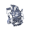
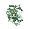
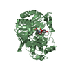
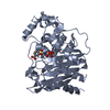
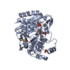
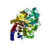
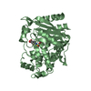
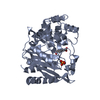
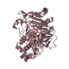
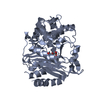
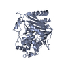
- PDB-1bls: CRYSTALLOGRAPHIC STRUCTURE OF A PHOSPHONATE DERIVATIVE OF THE ENT... -
+ Open data

- Basic information
| Entry | Database: PDB / ID: 1bls | ||||||
|---|---|---|---|---|---|---|---|
| Title | CRYSTALLOGRAPHIC STRUCTURE OF A PHOSPHONATE DERIVATIVE OF THE ENTEROBACTER CLOACAE P99 CEPHALOSPORINASE: MECHANISTIC INTERPRETATION OF A BETA-LACTAMASE TRANSITION STATE ANALOG | ||||||
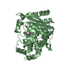
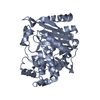
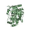
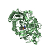
 Components Components | BETA-LACTAMASE | ||||||
 Keywords Keywords | HYDROLASE (ACTING IN CYCLIC AMIDES) | ||||||
| Function / homology |  Function and homology information Function and homology informationantibiotic catabolic process / beta-lactamase activity / beta-lactamase / outer membrane-bounded periplasmic space / response to antibiotic Similarity search - Function | ||||||
| Biological species |  Enterobacter cloacae (bacteria) Enterobacter cloacae (bacteria) | ||||||
| Method |  X-RAY DIFFRACTION / Resolution: 2.3 Å X-RAY DIFFRACTION / Resolution: 2.3 Å | ||||||
 Authors Authors | Knox, J.R. / Moews, P.C. / Lobkovsky, E. | ||||||
 Citation Citation | Journal: Biochemistry / Year: 1994 Title: Crystallographic structure of a phosphonate derivative of the Enterobacter cloacae P99 cephalosporinase: mechanistic interpretation of a beta-lactamase transition-state analog. Authors: Lobkovsky, E. / Billings, E.M. / Moews, P.C. / Rahil, J. / Pratt, R.F. / Knox, J.R. #1: Journal: Proc.Natl.Acad.Sci.USA / Year: 1993Title: Evolution of an Enzyme Activity: Crystallographic Structure at 2 Angstroms Resolution of the Cephalosporinase from the Ampc Gene of Enterobacter Cloacae P99 and Comparison with a Class a Penicillinase Authors: Lobkovsky, E. / Moews, P.C. / Liu, H. / Zhao, H. / Frere, J.M. / Knox, J.R. | ||||||
| History |
|
- Structure visualization
| Structure viewer | Molecule: MolmilJmol/JSmol |
|---|
- Downloads & links
-Download
| PDBx/mmCIF format | 1bls.cif.gz | 164.3 KB | Display | PDBx/mmCIF format |
|---|---|---|---|---|
| PDB format | pdb1bls.ent.gz | 129.7 KB | Display | PDB format |
| PDBx/mmJSON format | 1bls.json.gz | Tree view | PDBx/mmJSON format | |
| Others |  Other downloads Other downloads |
-Validation report
| Arichive directory | https://data.pdbj.org/pub/pdb/validation_reports/bl/1blsftp://data.pdbj.org/pub/pdb/validation_reports/bl/1bls | HTTPS FTP |
|---|
-Related structure data


-Links
 PDBj
PDBj
- Assembly
| Deposited unit | 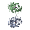
| ||||||||
|---|---|---|---|---|---|---|---|---|---|
| 1 | 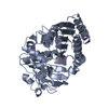
| ||||||||
| 2 | 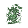
| ||||||||
| Unit cell |
| ||||||||
| Noncrystallographic symmetry (NCS) | NCS oper: (Code: given Matrix: (-0.999993, -0.000825, -0.003734), Vector: Details | THE *MTRIX* RECORDS BELOW MAY BE USED TO GENERATE APPROXIMATE COORDINATES OF MOLECULE B WHEN APPLIED TO MOLECULE A. | |
-Components
| #1: Protein | Mass: 39269.754 Da / Num. of mol.: 2 Source method: isolated from a genetically manipulated source Source: (gene. exp.) Enterobacter cloacae (bacteria) / References: UniProt: P05364, beta-lactamase#2: Chemical |   Mass: 339.067 Da / Num. of mol.: 2 / Source method: obtained synthetically / Formula: C9H11INO3P Mass: 339.067 Da / Num. of mol.: 2 / Source method: obtained synthetically / Formula: C9H11INO3P#3: Water | ChemComp-HOH / |  Mass: 18.015 Da / Num. of mol.: 789 / Source method: isolated from a natural source / Formula: H2O Mass: 18.015 Da / Num. of mol.: 789 / Source method: isolated from a natural source / Formula: H2OCompound details | TURN INFORMATION IS EASILY OBTAINED USING THE DSSP ANALYSIS PROGRAM BY KABSCH AND SANDER, ...TURN INFORMATIO | Has protein modification | Y | |
|---|
-Experimental details
-Experiment
| Experiment | Method: X-RAY DIFFRACTION |
|---|
- Sample preparation
| Crystal | Density Matthews: 2.36 Å3/Da / Density % sol: 47.82 % | |||||||||||||||
|---|---|---|---|---|---|---|---|---|---|---|---|---|---|---|---|---|
| Crystal grow | *PLUS pH: 6.5 / Method: unknown | |||||||||||||||
| Components of the solutions | *PLUS
|
-Data collection
| Radiation | Scattering type: x-ray |
|---|---|
| Radiation wavelength | Relative weight: 1 |
| Reflection | *PLUS Num. obs: 28780 / % possible obs: 91 % / Num. measured all: 82494 / Rmerge(I) obs: 0.044 |
| Reflection shell | *PLUS Highest resolution: 2.32 Å / Lowest resolution: 2.47 Å / % possible obs: 76 % / Num. unique obs: 7904 / Rmerge(I) obs: 0.158 |
- Processing
| Software | Name: PROLSQ / Classification: refinement | |||||||||||||||||||||||||||||||||||||||||||||||||||||||||||||||
|---|---|---|---|---|---|---|---|---|---|---|---|---|---|---|---|---|---|---|---|---|---|---|---|---|---|---|---|---|---|---|---|---|---|---|---|---|---|---|---|---|---|---|---|---|---|---|---|---|---|---|---|---|---|---|---|---|---|---|---|---|---|---|---|---|
| Refinement | Resolution: 2.3→8 Å /
| |||||||||||||||||||||||||||||||||||||||||||||||||||||||||||||||
| Refinement step | Cycle: LAST / Resolution: 2.3→8 Å
| |||||||||||||||||||||||||||||||||||||||||||||||||||||||||||||||
| Refine LS restraints |
| |||||||||||||||||||||||||||||||||||||||||||||||||||||||||||||||
| Refinement | *PLUS Highest resolution: 2.3 Å / Rfactor Rwork: 0.175 | |||||||||||||||||||||||||||||||||||||||||||||||||||||||||||||||
| Solvent computation | *PLUS | |||||||||||||||||||||||||||||||||||||||||||||||||||||||||||||||
| Displacement parameters | *PLUS Biso mean: 23 Å2 | |||||||||||||||||||||||||||||||||||||||||||||||||||||||||||||||
| Refine LS restraints | *PLUS
|
