 Movie
Movie Controller

[English] 日本語
 Yorodumi
- PDB-6g9e: Crystal structure of immunomodulatory active chitinase from Trich... -
+ Open data

- Basic information
| Entry | Database: PDB / ID: 6g9e | ||||||
|---|---|---|---|---|---|---|---|
| Title | Crystal structure of immunomodulatory active chitinase from Trichuris suis - TsES1 - 6 molecules in ASU | ||||||
 Components Components | Immunomodulatory active chitinase | ||||||
 Keywords Keywords | HYDROLASE / TsES1 / immunomodulatory potential / chitinase / Eukaryotic protein expression / LEXSY / secretion / dimer / intramolecular disulphide bridge | ||||||
| Function / homology |  Function and homology information Function and homology informationchitinase activity / chitin catabolic process / chitin binding / carbohydrate metabolic process / extracellular region Similarity search - Function | ||||||
| Biological species |  Trichuris suis (pig whipworm) Trichuris suis (pig whipworm) | ||||||
| Method |  X-RAY DIFFRACTION / SYNCHROTRON / MOLECULAR REPLACEMENT / Resolution: 2.69 Å X-RAY DIFFRACTION / SYNCHROTRON / MOLECULAR REPLACEMENT / Resolution: 2.69 Å | ||||||
 Authors Authors | Malecki, P.H. / Balster, K. / Hartmann, S. / Weiss, M.S. / Heinemann, U. | ||||||
 Citation Citation | Journal: J Immunol Res / Year: 2021 Title: A Helminth-Derived Chitinase Structurally Similar to Mammalian Chitinase Displays Immunomodulatory Properties in Inflammatory Lung Disease. Authors: Ebner, F. / Lindner, K. / Janek, K. / Niewienda, A. / Malecki, P.H. / Weiss, M.S. / Sutherland, T.E. / Heuser, A. / Kuhl, A.A. / Zentek, J. / Hofmann, A. / Hartmann, S. | ||||||
| History |
|
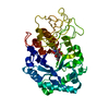
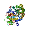
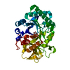
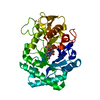
- Structure visualization
| Structure viewer | Molecule: MolmilJmol/JSmol |
|---|
- Downloads & links
-Download
| PDBx/mmCIF format | 6g9e.cif.gz | 895.7 KB | Display | PDBx/mmCIF format |
|---|---|---|---|---|
| PDB format | pdb6g9e.ent.gz | 746.5 KB | Display | PDB format |
| PDBx/mmJSON format | 6g9e.json.gz | Tree view | PDBx/mmJSON format | |
| Others |  Other downloads Other downloads |
-Validation report
| Arichive directory | https://data.pdbj.org/pub/pdb/validation_reports/g9/6g9eftp://data.pdbj.org/pub/pdb/validation_reports/g9/6g9e | HTTPS FTP |
|---|
-Related structure data
| Related structure data |  6g9cC 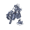 5hbfS S: Starting model for refinement C: citing same article ( |
|---|---|
| Similar structure data |


-Links
 PDBj
PDBj- Assembly
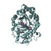
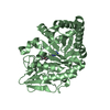
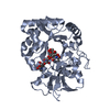
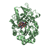
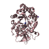
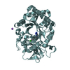
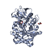
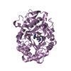
| Deposited unit | 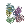
| ||||||||
|---|---|---|---|---|---|---|---|---|---|
| 1 | 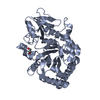
| ||||||||
| 2 | 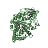
| ||||||||
| 3 | 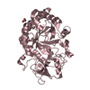
| ||||||||
| 4 | 
| ||||||||
| 5 | 
| ||||||||
| 6 | 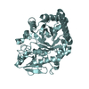
| ||||||||
| Unit cell |
| ||||||||
| Components on special symmetry positions |
|
-Components
| #1: Protein | Mass: 55545.691 Da / Num. of mol.: 6 / Fragment: TsES1 Source method: isolated from a genetically manipulated source Source: (gene. exp.) Trichuris suis (pig whipworm) / Gene: M513_10624 / Production host:  Leishmania tarentolae (eukaryote) / References: UniProt: A0A085LU44 Leishmania tarentolae (eukaryote) / References: UniProt: A0A085LU44#2: Chemical | ChemComp-EDO /   Mass: 62.068 Da / Num. of mol.: 5 / Source method: obtained synthetically / Formula: C2H6O2 Mass: 62.068 Da / Num. of mol.: 5 / Source method: obtained synthetically / Formula: C2H6O2#3: Water | ChemComp-HOH / |  Mass: 18.015 Da / Num. of mol.: 453 / Source method: isolated from a natural source / Formula: H2O Mass: 18.015 Da / Num. of mol.: 453 / Source method: isolated from a natural source / Formula: H2OHas protein modification | Y | |
|---|
-Experimental details
-Experiment
| Experiment | Method: X-RAY DIFFRACTION / Number of used crystals: 1 |
|---|
- Sample preparation
| Crystal | Density Matthews: 4.09 Å3/Da / Density % sol: 69.94 % |
|---|---|
| Crystal grow | Temperature: 293 K / Method: vapor diffusion / pH: 7 Details: 100mM Tris, 200mM Magnesium chloride hexahydrate, 10% PEG 8000 |
-Data collection
| Diffraction | Mean temperature: 100 K | ||||||||||||||||||||||||
|---|---|---|---|---|---|---|---|---|---|---|---|---|---|---|---|---|---|---|---|---|---|---|---|---|---|
| Diffraction source | Source: SYNCHROTRON / Site: BESSY  / Beamline: 14.1 / Wavelength: 0.9184 Å / Beamline: 14.1 / Wavelength: 0.9184 Å | ||||||||||||||||||||||||
| Detector | Type: DECTRIS PILATUS 6M / Detector: PIXEL / Date: Aug 3, 2017 | ||||||||||||||||||||||||
| Radiation | Monochromator: DOUBLE CRYSTAL / Protocol: SINGLE WAVELENGTH / Monochromatic (M) / Laue (L): M / Scattering type: x-ray | ||||||||||||||||||||||||
| Radiation wavelength | Wavelength: 0.9184 Å / Relative weight: 1 | ||||||||||||||||||||||||
| Reflection | Resolution: 2.69→45.61 Å / Num. obs: 110673 / % possible obs: 99 % / Redundancy: 3.9 % / Biso Wilson estimate: 37.83 Å2 / CC1/2: 0.967 / Rmerge(I) obs: 0.267 / Rpim(I) all: 0.155 / Rrim(I) all: 0.31 / Net I/σ(I): 4.2 | ||||||||||||||||||||||||
| Reflection shell | Diffraction-ID: 1
|
- Processing
| Software |
| ||||||||||||||||||||||||||||||||||||||||||||||||||||||||||||||||||||||||
|---|---|---|---|---|---|---|---|---|---|---|---|---|---|---|---|---|---|---|---|---|---|---|---|---|---|---|---|---|---|---|---|---|---|---|---|---|---|---|---|---|---|---|---|---|---|---|---|---|---|---|---|---|---|---|---|---|---|---|---|---|---|---|---|---|---|---|---|---|---|---|---|---|---|
| Refinement | Method to determine structure: MOLECULAR REPLACEMENT Starting model: 5hbf Resolution: 2.69→45.61 Å / SU ML: 0.39 / Cross valid method: THROUGHOUT / σ(F): 1.34 / Phase error: 25.44
| ||||||||||||||||||||||||||||||||||||||||||||||||||||||||||||||||||||||||
| Solvent computation | Shrinkage radii: 0.9 Å / VDW probe radii: 1.11 Å | ||||||||||||||||||||||||||||||||||||||||||||||||||||||||||||||||||||||||
| Displacement parameters | Biso max: 109.97 Å2 / Biso mean: 43.0582 Å2 / Biso min: 16.8 Å2 | ||||||||||||||||||||||||||||||||||||||||||||||||||||||||||||||||||||||||
| Refinement step | Cycle: final / Resolution: 2.69→45.61 Å
| ||||||||||||||||||||||||||||||||||||||||||||||||||||||||||||||||||||||||
| LS refinement shell | Refine-ID: X-RAY DIFFRACTION / Rfactor Rfree error: 0
| ||||||||||||||||||||||||||||||||||||||||||||||||||||||||||||||||||||||||
| Refinement TLS params. | Method: refined / Origin x: 12.3924 Å / Origin y: 5.3288 Å / Origin z: 130.6447 Å
| ||||||||||||||||||||||||||||||||||||||||||||||||||||||||||||||||||||||||
| Refinement TLS group |
|
