 Movie
Movie Controller

[English] 日本語
 Yorodumi
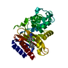
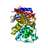
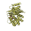
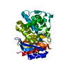
- PDB-1pzp: TEM-1 Beta-Lactamase in Complex with a Novel, Core-Disrupting, Al... -
+ Open data

- Basic information
| Entry | Database: PDB / ID: 1pzp | ||||||
|---|---|---|---|---|---|---|---|
| Title | TEM-1 Beta-Lactamase in Complex with a Novel, Core-Disrupting, Allosteric Inhibitor | ||||||
 Components Components | Beta-lactamase TEM | ||||||
 Keywords Keywords | HYDROLASE / beta-lactamase / novel allosteric inhibitor / core-disruption | ||||||
| Function / homology |  Function and homology information Function and homology informationAntimicrobial resistance / beta-lactam antibiotic catabolic process / beta-lactamase activity / beta-lactamase / response to antibiotic Similarity search - Function | ||||||
| Biological species |  | ||||||
| Method |  X-RAY DIFFRACTION / SYNCHROTRON / MOLECULAR REPLACEMENT / Resolution: 1.45 Å X-RAY DIFFRACTION / SYNCHROTRON / MOLECULAR REPLACEMENT / Resolution: 1.45 Å | ||||||
 Authors Authors | Horn, J.R. / Shoichet, B.K. | ||||||
 Citation Citation | Journal: J.Mol.Biol. / Year: 2004 Title: Allosteric inhibition through core disruption. Authors: Horn, J.R. / Shoichet, B.K. | ||||||
| History |
|
- Structure visualization
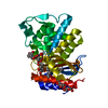
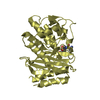
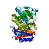
| Structure viewer | Molecule: MolmilJmol/JSmol |
|---|
- Downloads & links
-Download
| PDBx/mmCIF format | 1pzp.cif.gz | 70.8 KB | Display | PDBx/mmCIF format |
|---|---|---|---|---|
| PDB format | pdb1pzp.ent.gz | 51.3 KB | Display | PDB format |
| PDBx/mmJSON format | 1pzp.json.gz | Tree view | PDBx/mmJSON format | |
| Others |  Other downloads Other downloads |
-Validation report
| Arichive directory | https://data.pdbj.org/pub/pdb/validation_reports/pz/1pzpftp://data.pdbj.org/pub/pdb/validation_reports/pz/1pzp | HTTPS FTP |
|---|
-Related structure data


-Links
 PDBj
PDBj


- Assembly
| Deposited unit | 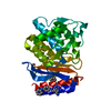
| ||||||||||
|---|---|---|---|---|---|---|---|---|---|---|---|
| 1 |
| ||||||||||
| Unit cell |
|
-Components
| #1: Protein | Mass: 28985.088 Da / Num. of mol.: 1 / Mutation: R100N Source method: isolated from a genetically manipulated source Source: (gene. exp.) | ||||
|---|---|---|---|---|---|
| #2: Chemical |   Mass: 303.321 Da / Num. of mol.: 2 / Source method: obtained synthetically / Formula: C16H13N7 Mass: 303.321 Da / Num. of mol.: 2 / Source method: obtained synthetically / Formula: C16H13N7#3: Water | ChemComp-HOH / |  Mass: 18.015 Da / Num. of mol.: 227 / Source method: isolated from a natural source / Formula: H2O Mass: 18.015 Da / Num. of mol.: 227 / Source method: isolated from a natural source / Formula: H2OHas protein modification | Y | |
-Experimental details
-Experiment
| Experiment | Method: X-RAY DIFFRACTION / Number of used crystals: 1 |
|---|
- Sample preparation
| Crystal | Density Matthews: 1.74 Å3/Da / Density % sol: 28.8 % |
|---|---|
| Crystal grow | Temperature: 295 K / Method: vapor diffusion, hanging drop / pH: 8.3 Details: potassium phosphate buffer, pH 8.3, VAPOR DIFFUSION, HANGING DROP, temperature 295K |
| Crystal grow | *PLUS pH: 8 / Method: other / Details: Wang, X., (2002) J.Mol.Biol., 320, 85. |
| Components of the solutions | *PLUS Conc.: 1.5 M / Common name: sodium phosphate / Details: pH8.0 |
-Data collection
| Diffraction | Mean temperature: 100 K |
|---|---|
| Diffraction source | Source: SYNCHROTRON / Site: APS  / Beamline: 5ID-B / Wavelength: 0.97014 Å / Beamline: 5ID-B / Wavelength: 0.97014 Å |
| Detector | Type: MARRESEARCH / Detector: CCD / Date: Mar 4, 2003 / Details: Mirrors |
| Radiation | Monochromator: graphite / Protocol: SINGLE WAVELENGTH / Monochromatic (M) / Laue (L): M / Scattering type: x-ray |
| Radiation wavelength | Wavelength: 0.97014 Å / Relative weight: 1 |
| Reflection | Resolution: 1.45→50 Å / Num. all: 40724 / Num. obs: 40562 / % possible obs: 99.6 % / Observed criterion σ(I): 3 / Redundancy: 4.9 % / Rmerge(I) obs: 0.06 / Net I/σ(I): 5.77 |
| Reflection shell | Resolution: 1.45→1.53 Å / Redundancy: 3.5 % / Rmerge(I) obs: 0.444 / Mean I/σ(I) obs: 1.6 / Num. unique all: 20643 / % possible all: 99.8 |
| Reflection | *PLUS Highest resolution: 1.47 Å / Lowest resolution: 20 Å / Num. obs: 38464 / % possible obs: 99.51 % / Rmerge(I) obs: 0.06 |
| Reflection shell | *PLUS Highest resolution: 1.47 Å / Lowest resolution: 1.58 Å / % possible obs: 99.8 % |
- Processing
| Software |
| ||||||||||||||||||||||||||||||||||||||||||||||||||||||||||||
|---|---|---|---|---|---|---|---|---|---|---|---|---|---|---|---|---|---|---|---|---|---|---|---|---|---|---|---|---|---|---|---|---|---|---|---|---|---|---|---|---|---|---|---|---|---|---|---|---|---|---|---|---|---|---|---|---|---|---|---|---|---|
| Refinement | Method to determine structure: MOLECULAR REPLACEMENT / Resolution: 1.45→50 Å / Cor.coef. Fo:Fc: 0.955 / Cor.coef. Fo:Fc free: 0.934 / SU B: 1.449 / SU ML: 0.057 / Cross valid method: THROUGHOUT / σ(F): 0 / σ(I): 0 / ESU R: 0.083 / ESU R Free: 0.089 / Stereochemistry target values: MAXIMUM LIKELIHOOD
| ||||||||||||||||||||||||||||||||||||||||||||||||||||||||||||
| Solvent computation | Ion probe radii: 0.8 Å / Shrinkage radii: 0.8 Å / VDW probe radii: 1.4 Å / Solvent model: BABINET MODEL WITH MASK | ||||||||||||||||||||||||||||||||||||||||||||||||||||||||||||
| Displacement parameters | Biso mean: 21.786 Å2
| ||||||||||||||||||||||||||||||||||||||||||||||||||||||||||||
| Refine analyze |
| ||||||||||||||||||||||||||||||||||||||||||||||||||||||||||||
| Refinement step | Cycle: LAST / Resolution: 1.45→50 Å
| ||||||||||||||||||||||||||||||||||||||||||||||||||||||||||||
| Refine LS restraints |
| ||||||||||||||||||||||||||||||||||||||||||||||||||||||||||||
| LS refinement shell | Resolution: 1.45→1.488 Å / Total num. of bins used: 20
| ||||||||||||||||||||||||||||||||||||||||||||||||||||||||||||
| Refinement | *PLUS Highest resolution: 1.47 Å / Lowest resolution: 20 Å / % reflection Rfree: 5 % / Rfactor Rfree: 0.234 / Rfactor Rwork: 0.193 | ||||||||||||||||||||||||||||||||||||||||||||||||||||||||||||
| Solvent computation | *PLUS | ||||||||||||||||||||||||||||||||||||||||||||||||||||||||||||
| Displacement parameters | *PLUS | ||||||||||||||||||||||||||||||||||||||||||||||||||||||||||||
| Refine LS restraints | *PLUS
|
