 Movie
Movie Controller


+ Open data

- Basic information
| Entry | Database: PDB / ID: 5l2d | ||||||
|---|---|---|---|---|---|---|---|
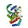
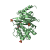
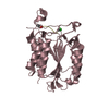
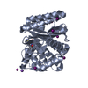
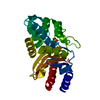
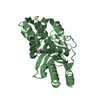
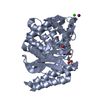
| Title | Streptococcal surface adhesin - CshA NR2 | ||||||
 Components Components | Surface-associated protein CshA | ||||||
 Keywords Keywords | CELL ADHESION / Streptococcus / adhesin | ||||||
| Function / homology |  Function and homology information Function and homology informationCshA domain / Surface adhesin CshA repetitive domain / GEVED domain / GEVED domain / YSIRK type signal peptide / YSIRK Gram-positive signal peptide / LPXTG cell wall anchor motif / Gram-positive cocci surface proteins LPxTG motif profile. / LPXTG cell wall anchor domain Similarity search - Domain/homology | ||||||
| Biological species |  Streptococcus gordonii (bacteria) Streptococcus gordonii (bacteria) | ||||||
| Method |  X-RAY DIFFRACTION / SYNCHROTRON / SAD / Resolution: 2.66 Å X-RAY DIFFRACTION / SYNCHROTRON / SAD / Resolution: 2.66 Å | ||||||
 Authors Authors | Back, C.R. / Race, P.R. / Jenkinson, H.F. | ||||||
 Citation Citation | Journal: J. Biol. Chem. / Year: 2017 Title: The Streptococcus gordonii Adhesin CshA Protein Binds Host Fibronectin via a Catch-Clamp Mechanism. Authors: Back, C.R. / Sztukowska, M.N. / Till, M. / Lamont, R.J. / Jenkinson, H.F. / Nobbs, A.H. / Race, P.R. | ||||||
| History |
|
- Structure visualization
| Structure viewer | Molecule: MolmilJmol/JSmol |
|---|
- Downloads & links
-Download
| PDBx/mmCIF format | 5l2d.cif.gz | 116.5 KB | Display | PDBx/mmCIF format |
|---|---|---|---|---|
| PDB format | pdb5l2d.ent.gz | 84.4 KB | Display | PDB format |
| PDBx/mmJSON format | 5l2d.json.gz | Tree view | PDBx/mmJSON format | |
| Others |  Other downloads Other downloads |
-Validation report
| Arichive directory | https://data.pdbj.org/pub/pdb/validation_reports/l2/5l2dftp://data.pdbj.org/pub/pdb/validation_reports/l2/5l2d | HTTPS FTP |
|---|
-Related structure data
| Similar structure data |
|---|


-Links
 PDBj
PDBj- Assembly
| Deposited unit | 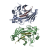
| ||||||||
|---|---|---|---|---|---|---|---|---|---|
| 1 | 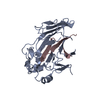
| ||||||||
| 2 | 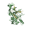
| ||||||||
| Unit cell |
|
-Components
| #1: Protein | Mass: 36776.012 Da / Num. of mol.: 4 / Fragment: UNP residues 223-540 Source method: isolated from a genetically manipulated source Source: (gene. exp.) Streptococcus gordonii (strain Challis / ATCC 35105 / BCRC 15272 / CH1 / DL1 / V288) (bacteria)Strain: Challis / ATCC 35105 / BCRC 15272 / CH1 / DL1 / V288 Gene: cshA, SGO_0854 / Production host: #2: Water | ChemComp-HOH / |  Mass: 18.015 Da / Num. of mol.: 2 / Source method: isolated from a natural source / Formula: H2O Mass: 18.015 Da / Num. of mol.: 2 / Source method: isolated from a natural source / Formula: H2O |
|---|
-Experimental details
-Experiment
| Experiment | Method: X-RAY DIFFRACTION / Number of used crystals: 1 Method details: The structure of CshA-NR2 was initially determined in space group P6222, with two molecules in the asymmetric unit, to 3.5 A resolution by the single wavelength anomalous dispersion ...Method details: The structure of CshA-NR2 was initially determined in space group P6222, with two molecules in the asymmetric unit, to 3.5 A resolution by the single wavelength anomalous dispersion (SAD) method as applied to selonomethionine (SeMet) labeled crystals of dimeric CshA-NR2. Identification of heavy atom sites and the resulting initial phase calculation was carried out, employing diffraction data collected from two different crystals of SeMet labeled dimeric CshA-NR2, which were merged together. The software located 18 Se sites. The output model was refined and used as a molecular replacement search model to elucidate a higher resolution CshA-NR2 structure (2.7 A), employing diffraction data collected from a crystal of unlabeled CshA-NR2 (also in space group P6222). |
|---|
- Sample preparation
| Crystal grow | Temperature: 291.15 K / Method: vapor diffusion, hanging drop Details: 0.2 M Na/K tartrate, MMT buffer [DL-malic acid, MES and Tris base in the molar ratios 1:2:2] (pH 5.0), 22% PEG 3350. |
|---|
-Data collection
| Diffraction | Mean temperature: 100 K |
|---|---|
| Diffraction source | Source: SYNCHROTRON / Site: Diamond  / Beamline: I04-1 / Wavelength: 0.92 Å / Beamline: I04-1 / Wavelength: 0.92 Å |
| Detector | Type: DECTRIS PILATUS 2M / Detector: PIXEL / Date: Sep 15, 2012 |
| Radiation | Protocol: SINGLE WAVELENGTH / Monochromatic (M) / Laue (L): M / Scattering type: x-ray |
| Radiation wavelength | Wavelength: 0.92 Å / Relative weight: 1 |
| Reflection | Resolution: 2.66→61.01 Å / Num. obs: 27284 / % possible obs: 100 % / Redundancy: 16.8 % / Rmerge(I) obs: 0.107 / Net I/σ(I): 19.8 |
| Reflection shell | Resolution: 2.66→2.8 Å / Redundancy: 16.6 % / Rmerge(I) obs: 1.477 / Mean I/σ(I) obs: 2.1 / % possible all: 100 |
- Processing
| Software |
| ||||||||||||||||||||||||||||||||||||||||||||||||||||||||||||||||||||||||||||||||||||||||||||||||||||||||||||||||||||||||||||||||||||||||||||||||||||||||||||||||||||||||||||||||||||||
|---|---|---|---|---|---|---|---|---|---|---|---|---|---|---|---|---|---|---|---|---|---|---|---|---|---|---|---|---|---|---|---|---|---|---|---|---|---|---|---|---|---|---|---|---|---|---|---|---|---|---|---|---|---|---|---|---|---|---|---|---|---|---|---|---|---|---|---|---|---|---|---|---|---|---|---|---|---|---|---|---|---|---|---|---|---|---|---|---|---|---|---|---|---|---|---|---|---|---|---|---|---|---|---|---|---|---|---|---|---|---|---|---|---|---|---|---|---|---|---|---|---|---|---|---|---|---|---|---|---|---|---|---|---|---|---|---|---|---|---|---|---|---|---|---|---|---|---|---|---|---|---|---|---|---|---|---|---|---|---|---|---|---|---|---|---|---|---|---|---|---|---|---|---|---|---|---|---|---|---|---|---|---|---|
| Refinement | Method to determine structure: SAD / Resolution: 2.66→61.01 Å / Cor.coef. Fo:Fc: 0.945 / Cor.coef. Fo:Fc free: 0.918 / SU B: 11.993 / SU ML: 0.234 / Cross valid method: THROUGHOUT / ESU R: 0.309 / ESU R Free: 0.267 / Stereochemistry target values: MAXIMUM LIKELIHOOD / Details: HYDROGENS HAVE BEEN ADDED IN THE RIDING POSITIONS
| ||||||||||||||||||||||||||||||||||||||||||||||||||||||||||||||||||||||||||||||||||||||||||||||||||||||||||||||||||||||||||||||||||||||||||||||||||||||||||||||||||||||||||||||||||||||
| Solvent computation | Ion probe radii: 0.8 Å / Shrinkage radii: 0.8 Å / VDW probe radii: 1.2 Å / Solvent model: MASK | ||||||||||||||||||||||||||||||||||||||||||||||||||||||||||||||||||||||||||||||||||||||||||||||||||||||||||||||||||||||||||||||||||||||||||||||||||||||||||||||||||||||||||||||||||||||
| Displacement parameters | Biso mean: 66.933 Å2
| ||||||||||||||||||||||||||||||||||||||||||||||||||||||||||||||||||||||||||||||||||||||||||||||||||||||||||||||||||||||||||||||||||||||||||||||||||||||||||||||||||||||||||||||||||||||
| Refinement step | Cycle: LAST / Resolution: 2.66→61.01 Å
| ||||||||||||||||||||||||||||||||||||||||||||||||||||||||||||||||||||||||||||||||||||||||||||||||||||||||||||||||||||||||||||||||||||||||||||||||||||||||||||||||||||||||||||||||||||||
| Refine LS restraints |
|