 Movie
Movie Controller

[English] 日本語
 Yorodumi
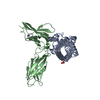
- PDB-3d48: Crystal structure of a prolactin receptor antagonist bound to the... -
+ Open data

- Basic information
| Entry | Database: PDB / ID: 3d48 | ||||||
|---|---|---|---|---|---|---|---|
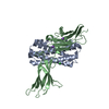
| Title | Crystal structure of a prolactin receptor antagonist bound to the extracellular domain of the prolactin receptor | ||||||
 Components Components |
| ||||||
 Keywords Keywords | Hormone/Hormone Receptor / Cytokine-Cytokine Receptor Complex / Four-helix Bundle / Glycoprotein / Hormone / Lactation / Secreted / Alternative splicing / Membrane / Receptor / Transmembrane / Hormone-Hormone Receptor COMPLEX | ||||||
| Function / homology |  Function and homology information Function and homology informationprolactin receptor activity / prolactin receptor binding / activation of transmembrane receptor protein tyrosine kinase activity / Developmental Lineage of Mammary Gland Alveolar Cells / prolactin signaling pathway / positive regulation of lactation / steroid biosynthetic process / activation of Janus kinase activity / cellular response to granulocyte macrophage colony-stimulating factor stimulus / mammary gland development ...prolactin receptor activity / prolactin receptor binding / activation of transmembrane receptor protein tyrosine kinase activity / Developmental Lineage of Mammary Gland Alveolar Cells / prolactin signaling pathway / positive regulation of lactation / steroid biosynthetic process / activation of Janus kinase activity / cellular response to granulocyte macrophage colony-stimulating factor stimulus / mammary gland development / negative regulation of endothelial cell proliferation / Prolactin receptor signaling / cytokine binding / peptide hormone binding / Growth hormone receptor signaling / lactation / embryo implantation / positive regulation of B cell proliferation / negative regulation of angiogenesis / endosome lumen / positive regulation of receptor signaling pathway via JAK-STAT / female pregnancy / hormone activity / response to nutrient levels / positive regulation of miRNA transcription / cytokine-mediated signaling pathway / positive regulation of cold-induced thermogenesis / positive regulation of canonical NF-kappaB signal transduction / signaling receptor complex / cell surface receptor signaling pathway / Amyloid fiber formation / external side of plasma membrane / positive regulation of cell population proliferation / lipid binding / negative regulation of apoptotic process / protein kinase binding / cell surface / : / extracellular region / metal ion binding / plasma membrane Similarity search - Function | ||||||
| Biological species |  Homo sapiens (human) Homo sapiens (human) | ||||||
| Method |  X-RAY DIFFRACTION / SYNCHROTRON / MOLECULAR REPLACEMENT / Resolution: 2.5 Å X-RAY DIFFRACTION / SYNCHROTRON / MOLECULAR REPLACEMENT / Resolution: 2.5 Å | ||||||
 Authors Authors | Svensson, L.A. / Breinholt, J. | ||||||
 Citation Citation | Journal: J.Biol.Chem. / Year: 2008 Title: Crystal structure of a prolactin receptor antagonist bound to the extracellular domain of the prolactin receptor Authors: Svensson, L.A. / Bondensgaard, K. / Norskov-Lauritsen, L. / Christensen, L. / Becker, P. / Andersen, M.D. / Maltesen, M.J. / Rand, K.D. / Breinholt, J. | ||||||
| History |
|
- Structure visualization
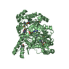
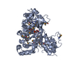
| Structure viewer | Molecule: MolmilJmol/JSmol |
|---|
- Downloads & links
-Download
| PDBx/mmCIF format | 3d48.cif.gz | 91.4 KB | Display | PDBx/mmCIF format |
|---|---|---|---|---|
| PDB format | pdb3d48.ent.gz | 68.1 KB | Display | PDB format |
| PDBx/mmJSON format | 3d48.json.gz | Tree view | PDBx/mmJSON format | |
| Others |  Other downloads Other downloads |
-Validation report
| Arichive directory | https://data.pdbj.org/pub/pdb/validation_reports/d4/3d48ftp://data.pdbj.org/pub/pdb/validation_reports/d4/3d48 | HTTPS FTP |
|---|
-Related structure data
| Related structure data |  1bp3S  1f6fS  1n9d  1rw5S S: Starting model for refinement |
|---|---|
| Similar structure data |


-Links
 PDBj
PDBj


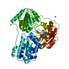
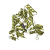
- Assembly
| Deposited unit | 
| ||||||||
|---|---|---|---|---|---|---|---|---|---|
| 1 |
| ||||||||
| Unit cell |
|
-Components
| #1: Protein | Mass: 21948.000 Da / Num. of mol.: 1 / Fragment: residues 12-199 / Mutation: Q12S, G129R Source method: isolated from a genetically manipulated source Source: (gene. exp.) Homo sapiens (human) / Gene: PRL / Plasmid: pET32-a(+) / Production host:  | ||||
|---|---|---|---|---|---|
| #2: Protein | Mass: 24529.850 Da / Num. of mol.: 1 / Fragment: Extracellular Domain Source method: isolated from a genetically manipulated source Source: (gene. exp.) Homo sapiens (human) / Gene: PRLR / Plasmid: pET39-b(+) / Production host: | ||||
| #3: Chemical | ChemComp-CO3 /   Mass: 60.009 Da / Num. of mol.: 4 / Source method: obtained synthetically / Formula: CO3 Mass: 60.009 Da / Num. of mol.: 4 / Source method: obtained synthetically / Formula: CO3#4: Water | ChemComp-HOH / |  Mass: 18.015 Da / Num. of mol.: 103 / Source method: isolated from a natural source / Formula: H2O Mass: 18.015 Da / Num. of mol.: 103 / Source method: isolated from a natural source / Formula: H2OHas protein modification | Y | |
-Experimental details
-Experiment
| Experiment | Method: X-RAY DIFFRACTION / Number of used crystals: 1 |
|---|
- Sample preparation
| Crystal | Density Matthews: 3.41 Å3/Da / Density % sol: 63.9 % |
|---|---|
| Crystal grow | Temperature: 297 K / Method: vapor diffusion, hanging drop / pH: 7.5 Details: 3.5M Sodium Chloride, 0.1M Hepes, pH 7.5, VAPOR DIFFUSION, HANGING DROP, temperature 297K |
-Data collection
| Diffraction | Mean temperature: 100 K |
|---|---|
| Diffraction source | Source: SYNCHROTRON / Site: MAX II  / Beamline: I911-3 / Wavelength: 1 Å / Beamline: I911-3 / Wavelength: 1 Å |
| Detector | Type: MARMOSAIC 225 mm CCD / Detector: CCD / Date: Apr 18, 2007 |
| Radiation | Monochromator: DOUBLE-CRYSTAL MONOCHROMATOR / Protocol: SINGLE WAVELENGTH / Monochromatic (M) / Laue (L): M / Scattering type: x-ray |
| Radiation wavelength | Wavelength: 1 Å / Relative weight: 1 |
| Reflection | Resolution: 2.5→20 Å / Num. all: 21763 / Num. obs: 21797 / % possible obs: 99.8 % / Observed criterion σ(F): -3 / Observed criterion σ(I): -3 / Redundancy: 11.2 % / Biso Wilson estimate: 46.4 Å2 / Rsym value: 0.083 / Net I/σ(I): 22.46 |
| Reflection shell | Resolution: 2.5→2.56 Å / Redundancy: 11.2 % / Mean I/σ(I) obs: 5.5 / Num. unique all: 21797 / Rsym value: 0.501 / % possible all: 100 |
- Processing
| Software |
| |||||||||||||||||||||||||||||||||||||||||||||||||||||||||||||||||||||||||||||||||||||||||||||||||||||||||||||||||||||||||||||||||||||||||||||||||||
|---|---|---|---|---|---|---|---|---|---|---|---|---|---|---|---|---|---|---|---|---|---|---|---|---|---|---|---|---|---|---|---|---|---|---|---|---|---|---|---|---|---|---|---|---|---|---|---|---|---|---|---|---|---|---|---|---|---|---|---|---|---|---|---|---|---|---|---|---|---|---|---|---|---|---|---|---|---|---|---|---|---|---|---|---|---|---|---|---|---|---|---|---|---|---|---|---|---|---|---|---|---|---|---|---|---|---|---|---|---|---|---|---|---|---|---|---|---|---|---|---|---|---|---|---|---|---|---|---|---|---|---|---|---|---|---|---|---|---|---|---|---|---|---|---|---|---|---|---|
| Refinement | Method to determine structure: MOLECULAR REPLACEMENT Starting model: FOR THE R CHAIN, PDB ENTRY 1BP3. FOR THE P CHAIN, A CORE OF A MODELER HOMOLOGY MODEL BASED ON PDB ENTRIES 1BP3, 1F6F, 1RW5 AND 1N9D. Resolution: 2.5→20 Å / Cor.coef. Fo:Fc: 0.928 / Cor.coef. Fo:Fc free: 0.863 / WRfactor Rfree: 0.29 / WRfactor Rwork: 0.212 / SU B: 9.77 / SU ML: 0.222 / Cross valid method: THROUGHOUT / σ(F): 0 / ESU R: 0.335 / ESU R Free: 0.289 / Stereochemistry target values: MAXIMUM LIKELIHOOD
| |||||||||||||||||||||||||||||||||||||||||||||||||||||||||||||||||||||||||||||||||||||||||||||||||||||||||||||||||||||||||||||||||||||||||||||||||||
| Solvent computation | Ion probe radii: 0.8 Å / Shrinkage radii: 0.8 Å / VDW probe radii: 1.2 Å / Solvent model: MASK | |||||||||||||||||||||||||||||||||||||||||||||||||||||||||||||||||||||||||||||||||||||||||||||||||||||||||||||||||||||||||||||||||||||||||||||||||||
| Displacement parameters | Biso mean: 46.307 Å2
| |||||||||||||||||||||||||||||||||||||||||||||||||||||||||||||||||||||||||||||||||||||||||||||||||||||||||||||||||||||||||||||||||||||||||||||||||||
| Refinement step | Cycle: LAST / Resolution: 2.5→20 Å
| |||||||||||||||||||||||||||||||||||||||||||||||||||||||||||||||||||||||||||||||||||||||||||||||||||||||||||||||||||||||||||||||||||||||||||||||||||
| Refine LS restraints |
| |||||||||||||||||||||||||||||||||||||||||||||||||||||||||||||||||||||||||||||||||||||||||||||||||||||||||||||||||||||||||||||||||||||||||||||||||||
| LS refinement shell | Refine-ID: X-RAY DIFFRACTION / Total num. of bins used: 20
|
