 Movie
Movie Controller

[English] 日本語
 Yorodumi
- PDB-1f6f: CRYSTAL STRUCTURE OF THE TERNARY COMPLEX BETWEEN OVINE PLACENTAL ... -
+ Open data

- Basic information
| Entry | Database: PDB / ID: 1f6f | ||||||
|---|---|---|---|---|---|---|---|
| Title | CRYSTAL STRUCTURE OF THE TERNARY COMPLEX BETWEEN OVINE PLACENTAL LACTOGEN AND THE EXTRACELLULAR DOMAIN OF THE RAT PROLACTIN RECEPTOR | ||||||
 Components Components |
| ||||||
 Keywords Keywords | HORMONE/GROWTH FACTOR/HORMONE RECEPTOR / 4-helical bundle / alpha helical bundle / ternary complex / FN III domains / beta sheet domains / cytokine-receptor complex / HORMONE-GROWTH FACTOR-HORMONE RECEPTOR COMPLEX | ||||||
| Function / homology |  Function and homology information Function and homology informationProlactin receptor signaling / prolactin receptor activity / prolactin receptor binding / regulation of epithelial cell differentiation / Growth hormone receptor signaling / prostate gland growth / mammary gland epithelial cell differentiation / positive regulation of lactation / mammary gland epithelium development / cellular response to granulocyte macrophage colony-stimulating factor stimulus ...Prolactin receptor signaling / prolactin receptor activity / prolactin receptor binding / regulation of epithelial cell differentiation / Growth hormone receptor signaling / prostate gland growth / mammary gland epithelial cell differentiation / positive regulation of lactation / mammary gland epithelium development / cellular response to granulocyte macrophage colony-stimulating factor stimulus / mammary gland development / mammary gland alveolus development / cytokine binding / peptide hormone binding / regulation of cell adhesion / cell surface receptor signaling pathway via JAK-STAT / lactation / response to bacterium / positive regulation of B cell proliferation / positive regulation of receptor signaling pathway via JAK-STAT / female pregnancy / hormone activity / response to nutrient levels / cytokine-mediated signaling pathway / positive regulation of cold-induced thermogenesis / signaling receptor complex / external side of plasma membrane / positive regulation of cell population proliferation / lipid binding / negative regulation of apoptotic process / protein kinase binding / cell surface / : / metal ion binding / plasma membrane Similarity search - Function | ||||||
| Biological species |  | ||||||
| Method |  X-RAY DIFFRACTION / SYNCHROTRON / Resolution: 2.3 Å X-RAY DIFFRACTION / SYNCHROTRON / Resolution: 2.3 Å | ||||||
 Authors Authors | Elkins, P.A. / Christinger, H.W. / Sandowski, Y. / Sakal, E. / Gertler, A. / De Vos, A.M. / Kossiakoff, A.A. | ||||||
 Citation Citation | Journal: Nat.Struct.Biol. / Year: 2000 Title: Ternary complex between placental lactogen and the extracellular domain of the prolactin receptor. Authors: Elkins, P.A. / Christinger, H.W. / Sandowski, Y. / Sakal, E. / Gertler, A. / de Vos, A.M. / Kossiakoff, A.A. #1: Journal: Acta Crystallogr.,Sect.D / Year: 1998Title: Crystallization of Ovine Placental Lactogen in a 1:2 Complex with the Extracellular Domain of the Rat Prolactin Receptor Authors: Christinger, H.W. / Elkins, P.A. / Sandowski, Y. / Sakal, E. / Gertler, A. / Kossiakoff, A.A. / De Vos, A.M. | ||||||
| History |
|
- Structure visualization
| Structure viewer | Molecule: MolmilJmol/JSmol |
|---|
- Downloads & links
-Download
| PDBx/mmCIF format | 1f6f.cif.gz | 124.6 KB | Display | PDBx/mmCIF format |
|---|---|---|---|---|
| PDB format | pdb1f6f.ent.gz | 98.4 KB | Display | PDB format |
| PDBx/mmJSON format | 1f6f.json.gz | Tree view | PDBx/mmJSON format | |
| Others |  Other downloads Other downloads |
-Validation report
| Arichive directory | https://data.pdbj.org/pub/pdb/validation_reports/f6/1f6fftp://data.pdbj.org/pub/pdb/validation_reports/f6/1f6f | HTTPS FTP |
|---|
-Related structure data
| Similar structure data |
|---|


-Links
 PDBj
PDBj


- Assembly
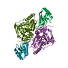
| Deposited unit | 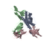
| ||||||||
|---|---|---|---|---|---|---|---|---|---|
| 1 |
| ||||||||
| Unit cell |
| ||||||||
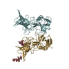
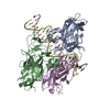
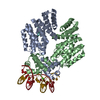
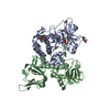
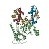
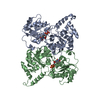
| Details | The biological assembly is a complex of the hormone (chain A) bound to 2 copies of the receptor (chains B and C). The copies of the receptor are not related by precise symmetry. |
-Components
| #1: Protein | Mass: 22601.643 Da / Num. of mol.: 1 Source method: isolated from a genetically manipulated source Source: (gene. exp.)  | ||||
|---|---|---|---|---|---|
| #2: Protein | Mass: 24584.494 Da / Num. of mol.: 2 Fragment: EXTRACELLULAR DOMAIN; N-TERMINAL FIBRONECTIN TYPE III DOMAINS Source method: isolated from a genetically manipulated source Source: (gene. exp.) #3: Water | ChemComp-HOH / |  Mass: 18.015 Da / Num. of mol.: 120 / Source method: isolated from a natural source / Formula: H2O Mass: 18.015 Da / Num. of mol.: 120 / Source method: isolated from a natural source / Formula: H2OHas protein modification | Y | |
-Experimental details
-Experiment
| Experiment | Method: X-RAY DIFFRACTION / Number of used crystals: 1 |
|---|
- Sample preparation
| Crystal | Density Matthews: 2.87 Å3/Da / Density % sol: 57.1 % | |||||||||||||||||||||||||
|---|---|---|---|---|---|---|---|---|---|---|---|---|---|---|---|---|---|---|---|---|---|---|---|---|---|---|
| Crystal grow | Temperature: 294 K / Method: vapor diffusion, hanging drop / pH: 5.6 Details: PEG 4000, isopropanol, MPD, MES, pH 5.6, VAPOR DIFFUSION, HANGING DROP, temperature 294K | |||||||||||||||||||||||||
| Crystal grow | *PLUS Details: Christinger, H.W., (1998) Acta Crystallogr., Sect.D, 54, 1408. | |||||||||||||||||||||||||
| Components of the solutions | *PLUS
|
-Data collection
| Diffraction source | Source: SYNCHROTRON / Site: CHESS  / Beamline: A1 / Beamline: A1 |
|---|---|
| Detector | Type: ADSC / Detector: CCD |
| Radiation | Protocol: SINGLE WAVELENGTH / Monochromatic (M) / Laue (L): M / Scattering type: x-ray |
| Radiation wavelength | Relative weight: 1 |
| Reflection | Resolution: 2.3→30 Å / Num. all: 36413 / Num. obs: 33034 / % possible obs: 90.7 % / Biso Wilson estimate: 47.9 Å2 / Rmerge(I) obs: 0.06 / Net I/σ(I): 16.2 |
| Reflection shell | Resolution: 2.3→30 Å / Rmerge(I) obs: 0.13 / Num. unique all: 2537 / % possible all: 70.4 |
| Reflection shell | *PLUS % possible obs: 70.4 % |
- Processing
| Software |
| |||||||||||||||||||||||||
|---|---|---|---|---|---|---|---|---|---|---|---|---|---|---|---|---|---|---|---|---|---|---|---|---|---|---|
| Refinement | Resolution: 2.3→30 Å / σ(F): 0.01 / Stereochemistry target values: Engh & Huber Details: Model bias was reduced by adding experimental phase information using cross-crystal averaging from a non-isomorphous mercury derivative.
| |||||||||||||||||||||||||
| Refinement step | Cycle: LAST / Resolution: 2.3→30 Å
| |||||||||||||||||||||||||
| Refine LS restraints |
|
