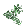
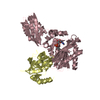
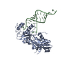
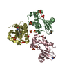
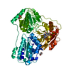
[English] 日本語
Yorodumi
- PDB-5dn8: 1.76 Angstrom Crystal Structure of GTP-binding Protein Der from C... -

+
Open data


ID or keywords:

Loading...

-
Basic information

Entry
Database: PDB / ID: 5dn8
Title1.76 Angstrom Crystal Structure of GTP-binding Protein Der from Coxiella burnetii in Complex with GDP.
ComponentsGTPase Der
KeywordsGTP-binding protein / GDP / Complex / Structural genomics / CSGID / Center for Structural Genomics of Infectious Diseases
Function / homology
Function and homology information


ribosomal large subunit assembly / ribosome binding / GTP binding
Similarity search - Function
GTP-binding protein EngA / EngA-type guanine nucleotide-binding (G) domain / EngA-type guanine nucleotide-binding (G) domain profile. / GTPase Der, C-terminal KH-domain-like / KH-domain-like of EngA bacterial GTPase enzymes, C-terminal / 50S ribosome-binding GTPase / GTP binding domain / K homology domain-like, alpha/beta / Small GTP-binding protein domain / P-loop containing nucleoside triphosphate hydrolase
Similarity search - Domain/homology
GUANOSINE-5'-DIPHOSPHATE / GTPase Der
Similarity search - Component
Biological speciesCoxiella burnetii (bacteria)
MethodX-RAY DIFFRACTION / SYNCHROTRON / MOLECULAR REPLACEMENT / Resolution: 1.76 Å
AuthorsMinasov, G. / Shuvalova, L. / Han, A. / Kim, H.-Y. / Grimshaw, S. / Kwon, K. / Anderson, W.F. / Center for Structural Genomics of Infectious Diseases (CSGID)
CitationJournal: To Be Published
Title: 1.76 Angstrom Crystal Structure of GTP-binding Protein Der from Coxiella burnetii in Complex with GDP.
Authors: Minasov, G. / Shuvalova, L. / Han, A. / Kim, H.-Y. / Grimshaw, S. / Kwon, K. / Anderson, W.F. / Center for Structural Genomics of Infectious Diseases (CSGID)
History
DepositionSep 9, 2015Deposition site: RCSB / Processing site: RCSB
Revision 1.0Oct 14, 2015Provider: repository / Type: Initial release
Revision 1.1Sep 27, 2023Group: Data collection / Database references ...Data collection / Database references / Derived calculations / Refinement description
Category: chem_comp_atom / chem_comp_bond ...chem_comp_atom / chem_comp_bond / database_2 / pdbx_initial_refinement_model / pdbx_prerelease_seq / pdbx_struct_oper_list
Item: _database_2.pdbx_DOI / _database_2.pdbx_database_accession / _pdbx_struct_oper_list.symmetry_operation

-
Structure visualization

Structure viewerMolecule:
MolmilJmol/JSmol

Downloads & links

-
Assembly

Deposited unit
A: GTPase Der
hetero molecules


Theoretical massNumber of molelcules
Total (without water)50,9373
Polymers50,0501
Non-polymers8862
Water5,423301
1


  • Idetical with deposited unit
  • defined by author&software
TypeNameSymmetry operationNumber
identity operation1_555x,y,z1
Buried area1250 Å2
ΔGint-9 kcal/mol
Surface area19830 Å2
MethodPISA
Unit cell
Length a, b, c (Å)44.275, 98.443, 107.968
Angle α, β, γ (deg.)90.00, 90.00, 90.00
Int Tables number19
Space group name H-MP212121

-
Components

#1: Protein GTPase Der / GTP-binding protein EngA


Mass: 50050.398 Da / Num. of mol.: 1
Source method: isolated from a genetically manipulated source
Source: (gene. exp.) Coxiella burnetii (strain RSA 493 / Nine Mile phase I) (bacteria)
Strain: RSA 493 / Nine Mile phase I / Gene: der, engA, CBU_1245 / Plasmid: pMCSG7 / Production host: ESCHERICHIA COLI (E. coli) / Strain (production host): BL21 magic / References: UniProt: Q83C83
#2: Chemical ChemComp-GDP / GUANOSINE-5'-DIPHOSPHATE


Type: RNA linking / Mass: 443.201 Da / Num. of mol.: 2 / Source method: obtained synthetically / Formula: C10H15N5O11P2 / Comment: GDP, energy-carrying molecule*YM
#3: Water ChemComp-HOH / water


Mass: 18.015 Da / Num. of mol.: 301 / Source method: isolated from a natural source / Formula: H2O

-
Experimental details

-
Experiment

ExperimentMethod: X-RAY DIFFRACTION

-
Sample preparation

CrystalDensity Matthews: 2.37 Å3/Da / Density % sol: 48.1 %
Crystal growTemperature: 295 K / Method: vapor diffusion, sitting drop / pH: 5.5
Details: Protein: 11.0 mg/ml, 0.25M Sodium chloride, 0.01M Tris-HCl pH 8.3, 10mM GDP, Screen: JSCG+ (H11), 0.2M Magnesium chloride, 0.1M Bis-Tris pH 5.5, 25% (w/v) PEG 3350.

-
Data collection

DiffractionMean temperature: 100 K
Diffraction sourceSource: SYNCHROTRON / Site: APS / Beamline: 21-ID-G / Wavelength: 0.97856 Å
DetectorType: MARMOSAIC 325 mm CCD / Detector: CCD / Date: Oct 2, 2014
RadiationMonochromator: C(111) / Protocol: SINGLE WAVELENGTH / Monochromatic (M) / Laue (L): M / Scattering type: x-ray
Radiation wavelengthWavelength: 0.97856 Å / Relative weight: 1
ReflectionResolution: 1.76→30 Å / Num. obs: 47308 / % possible obs: 99.7 % / Observed criterion σ(I): -3 / Redundancy: 7.2 % / Rmerge(I) obs: 0.051 / Rsym value: 0.051 / Net I/σ(I): 33.8
Reflection shellResolution: 1.76→1.79 Å / Redundancy: 7.3 % / Rmerge(I) obs: 0.537 / Mean I/σ(I) obs: 3.9 / % possible all: 100

-
Processing

Software
NameVersionClassification
REFMAC5.8.0073refinement
BLU-MAXdata collection
HKL-2000data scaling
PHASERphasing
HKL-2000data reduction
RefinementMethod to determine structure: MOLECULAR REPLACEMENT
Starting model: 2HJC

2hjc
PDB Unreleased entry


Resolution: 1.76→29.05 Å / Cor.coef. Fo:Fc: 0.966 / Cor.coef. Fo:Fc free: 0.951 / SU B: 3.995 / SU ML: 0.069 / Cross valid method: THROUGHOUT / ESU R: 0.113 / ESU R Free: 0.109 / Stereochemistry target values: MAXIMUM LIKELIHOOD / Details: HYDROGENS HAVE BEEN ADDED IN THE RIDING POSITIONS
RfactorNum. reflection% reflectionSelection details
Rfree0.2044 2386 5.1 %RANDOM
Rwork0.17128 ---
obs0.17295 44856 99.21 %-
Solvent computationIon probe radii: 0.8 Å / Shrinkage radii: 0.8 Å / VDW probe radii: 1.2 Å / Solvent model: MASK
Displacement parametersBiso mean: 39.181 Å2
Baniso -1Baniso -2Baniso -3
1-0.61 Å20 Å20 Å2
2---0.85 Å20 Å2
3---0.24 Å2
Refinement stepCycle: 1 / Resolution: 1.76→29.05 Å
ProteinNucleic acidLigandSolventTotal
Num. atoms3187 0 56 301 3544
Refine LS restraints
Refine-IDTypeDev idealDev ideal targetNumber
X-RAY DIFFRACTIONr_bond_refined_d0.0090.0193705
X-RAY DIFFRACTIONr_bond_other_d0.0010.023679
X-RAY DIFFRACTIONr_angle_refined_deg1.4731.9865059
X-RAY DIFFRACTIONr_angle_other_deg0.74638428
X-RAY DIFFRACTIONr_dihedral_angle_1_deg3.2175477
X-RAY DIFFRACTIONr_dihedral_angle_2_deg27.98922.84169
X-RAY DIFFRACTIONr_dihedral_angle_3_deg10.23915673
X-RAY DIFFRACTIONr_dihedral_angle_4_deg12.3191542
X-RAY DIFFRACTIONr_chiral_restr0.0840.2571
X-RAY DIFFRACTIONr_gen_planes_refined0.020.024301
X-RAY DIFFRACTIONr_gen_planes_other0.0160.02889
X-RAY DIFFRACTIONr_nbd_refined
X-RAY DIFFRACTIONr_nbd_other
X-RAY DIFFRACTIONr_nbtor_refined
X-RAY DIFFRACTIONr_nbtor_other
X-RAY DIFFRACTIONr_xyhbond_nbd_refined
X-RAY DIFFRACTIONr_xyhbond_nbd_other
X-RAY DIFFRACTIONr_metal_ion_refined
X-RAY DIFFRACTIONr_metal_ion_other
X-RAY DIFFRACTIONr_symmetry_vdw_refined
X-RAY DIFFRACTIONr_symmetry_vdw_other
X-RAY DIFFRACTIONr_symmetry_hbond_refined
X-RAY DIFFRACTIONr_symmetry_hbond_other
X-RAY DIFFRACTIONr_symmetry_metal_ion_refined
X-RAY DIFFRACTIONr_symmetry_metal_ion_other
X-RAY DIFFRACTIONr_mcbond_it2.1142.5161827
X-RAY DIFFRACTIONr_mcbond_other2.1082.5141826
X-RAY DIFFRACTIONr_mcangle_it3.2183.7412331
X-RAY DIFFRACTIONr_mcangle_other3.2173.7432332
X-RAY DIFFRACTIONr_scbond_it2.8122.9591878
X-RAY DIFFRACTIONr_scbond_other2.8162.9611870
X-RAY DIFFRACTIONr_scangle_it
X-RAY DIFFRACTIONr_scangle_other4.4764.2872716
X-RAY DIFFRACTIONr_long_range_B_refined7.46721.1974205
X-RAY DIFFRACTIONr_long_range_B_other7.34220.5864087
X-RAY DIFFRACTIONr_rigid_bond_restr
X-RAY DIFFRACTIONr_sphericity_free
X-RAY DIFFRACTIONr_sphericity_bonded
LS refinement shellResolution: 1.761→1.806 Å
RfactorNum. reflection% reflection
Rfree0.272 152 5 %
Rwork0.223 3055 -
obs--93.55 %
Refinement TLS params.

Method: refined / Refine-ID: X-RAY DIFFRACTION

IDL112)L122)L132)L222)L232)L332)S11 (Å °)S12 (Å °)S13 (Å °)S21 (Å °)S22 (Å °)S23 (Å °)S31 (Å °)S32 (Å °)S33 (Å °)T112)T122)T132)T222)T232)T332)Origin x (Å)Origin y (Å)Origin z (Å)
11.19240.14940.07972.38580.44922.053-0.0093-0.0013-0.14940.14430.044-0.16530.185-0.0322-0.03460.0326-0.0084-0.01160.0524-0.02760.04667.6717-6.1517-7.7096
20.0329-0.08310.39160.4094-1.86648.59660.0837-0.03350.01530.01430.18890.03770.0442-0.7555-0.27260.4037-0.01330.00980.272-0.02830.27-2.7291-14.494-28.9591
32.13051.0390.14372.03890.1031.26390.0612-0.09130.04470.149-0.0444-0.11850.0280.0842-0.01680.05310.02380.01570.0360.01330.02997.551-35.0782-25.8203
42.5213-0.73740.68782.943-1.21442.54860.0512-0.0731-0.0065-0.06540.05520.28190.0522-0.2849-0.10640.0649-0.03250.01980.13570.03690.0611-0.2827-11.921-46.0517
Refinement TLS group
IDRefine-IDRefine TLS-IDAuth asym-IDAuth seq-ID
1X-RAY DIFFRACTION1A0 - 23
2X-RAY DIFFRACTION1A41 - 164
3X-RAY DIFFRACTION2A24 - 40
4X-RAY DIFFRACTION3A173 - 350
5X-RAY DIFFRACTION4A351 - 434

+
About Yorodumi

-
News

-
Feb 9, 2022. New format data for meta-information of EMDB entries

New format data for meta-information of EMDB entries

  • Version 3 of the EMDB header file is now the official format.
  • The previous official version 1.9 will be removed from the archive.

Related info.:EMDB header

External links:wwPDB to switch to version 3 of the EMDB data model

-
Aug 12, 2020. Covid-19 info

Covid-19 info

URL: https://pdbjlvh1.pdbj.org/emnavi/covid19.php

New page: Covid-19 featured information page in EM Navigator.

Related info.:Covid-19 info / Mar 5, 2020. Novel coronavirus structure data

+
Mar 5, 2020. Novel coronavirus structure data

Novel coronavirus structure data

Related info.:Yorodumi Speices / Aug 12, 2020. Covid-19 info

External links:COVID-19 featured content - PDBj / Molecule of the Month (242):Coronavirus Proteases

+
Jan 31, 2019. EMDB accession codes are about to change! (news from PDBe EMDB page)

EMDB accession codes are about to change! (news from PDBe EMDB page)

  • The allocation of 4 digits for EMDB accession codes will soon come to an end. Whilst these codes will remain in use, new EMDB accession codes will include an additional digit and will expand incrementally as the available range of codes is exhausted. The current 4-digit format prefixed with “EMD-” (i.e. EMD-XXXX) will advance to a 5-digit format (i.e. EMD-XXXXX), and so on. It is currently estimated that the 4-digit codes will be depleted around Spring 2019, at which point the 5-digit format will come into force.
  • The EM Navigator/Yorodumi systems omit the EMD- prefix.

Related info.:Q: What is EMD? / ID/Accession-code notation in Yorodumi/EM Navigator

External links:EMDB Accession Codes are Changing Soon! / Contact to PDBj

+
Jul 12, 2017. Major update of PDB

Major update of PDB

  • wwPDB released updated PDB data conforming to the new PDBx/mmCIF dictionary.
  • This is a major update changing the version number from 4 to 5, and with Remediation, in which all the entries are updated.
  • In this update, many items about electron microscopy experimental information are reorganized (e.g. em_software).
  • Now, EM Navigator and Yorodumi are based on the updated data.

External links:wwPDB Remediation / Enriched Model Files Conforming to OneDep Data Standards Now Available in the PDB FTP Archive

-
Yorodumi

Thousand views of thousand structures

  • Yorodumi is a browser for structure data from EMDB, PDB, SASBDB, etc.
  • This page is also the successor to EM Navigator detail page, and also detail information page/front-end page for Omokage search.
  • The word "yorodu" (or yorozu) is an old Japanese word meaning "ten thousand". "mi" (miru) is to see.

Related info.:EMDB / PDB / SASBDB / Comparison of 3 databanks / Yorodumi Search / Aug 31, 2016. New EM Navigator & Yorodumi / Yorodumi Papers / Jmol/JSmol / Function and homology information / Changes in new EM Navigator and Yorodumi

Read more