 Movie
Movie Controller

[English] 日本語
 Yorodumi
- PDB-3d2u: Structure of UL18, a Peptide-Binding Viral MHC Mimic, Bound to a ... -
+ Open data

- Basic information
| Entry | Database: PDB / ID: 3d2u | ||||||
|---|---|---|---|---|---|---|---|
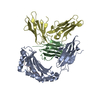
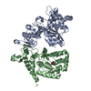
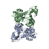
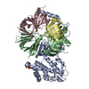
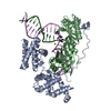
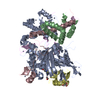
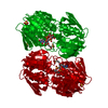
| Title | Structure of UL18, a Peptide-Binding Viral MHC Mimic, Bound to a Host Inhibitory Receptor | ||||||
 Components Components |
| ||||||
 Keywords Keywords | IMMUNE SYSTEM / MHC class I homolog / Disease mutation / Glycation / Glycoprotein / Immune response / Immunoglobulin domain / MHC I / Pyrrolidone carboxylic acid / Secreted / Membrane / Phosphoprotein / Receptor / Transmembrane | ||||||
| Function / homology |  Function and homology information Function and homology informationsymbiont-mediated perturbation of host natural killer cell mediated immune response / HLA-A specific inhibitory MHC class I receptor activity / : / negative regulation of serotonin secretion / MHC class Ib protein complex binding / HLA-B specific inhibitory MHC class I receptor activity / inhibitory MHC class I receptor activity / negative regulation of dendritic cell differentiation / Fc receptor mediated inhibitory signaling pathway / immune response-inhibiting cell surface receptor signaling pathway ...symbiont-mediated perturbation of host natural killer cell mediated immune response / HLA-A specific inhibitory MHC class I receptor activity / : / negative regulation of serotonin secretion / MHC class Ib protein complex binding / HLA-B specific inhibitory MHC class I receptor activity / inhibitory MHC class I receptor activity / negative regulation of dendritic cell differentiation / Fc receptor mediated inhibitory signaling pathway / immune response-inhibiting cell surface receptor signaling pathway / MHC class Ib receptor activity / MHC class Ib protein binding / negative regulation of T cell mediated cytotoxicity / immune response-regulating signaling pathway / negative regulation of CD8-positive, alpha-beta T cell activation / negative regulation of transforming growth factor beta production / MHC class I receptor activity / negative regulation of cytokine production involved in immune response / positive regulation of norepinephrine uptake / dendritic cell differentiation / interleukin-10-mediated signaling pathway / negative regulation of osteoclast development / negative regulation of interleukin-12 production / protein phosphatase 1 binding / negative regulation of T cell activation via T cell receptor contact with antigen bound to MHC molecule on antigen presenting cell / bBAF complex / cellular response to cytochalasin B / Formation of the embryonic stem cell BAF (esBAF) complex / negative regulation of alpha-beta T cell activation / npBAF complex / brahma complex / nBAF complex / Formation of the canonical BAF (cBAF) complex / regulation of transepithelial transport / negative regulation of dendritic cell apoptotic process / morphogenesis of a polarized epithelium / Formation of the polybromo-BAF (pBAF) complex / Formation of neuronal progenitor and neuronal BAF (npBAF and nBAF) / structural constituent of postsynaptic actin cytoskeleton / Formation of annular gap junctions / Formation of the dystrophin-glycoprotein complex (DGC) / GBAF complex / Gap junction degradation / negative regulation of endocytosis / Formation of the non-canonical BAF (ncBAF) complex / protein localization to adherens junction / regulation of G0 to G1 transition / Cell-extracellular matrix interactions / negative regulation of interferon-beta production / dense body / Folding of actin by CCT/TriC / Tat protein binding / negative regulation of mononuclear cell proliferation / postsynaptic actin cytoskeleton / negative regulation of natural killer cell mediated cytotoxicity / RSC-type complex / Regulation of CDH1 Function / regulation of double-strand break repair / Prefoldin mediated transfer of substrate to CCT/TriC / regulation of nucleotide-excision repair / Adherens junctions interactions / adherens junction assembly / RHOF GTPase cycle / apical protein localization / T cell proliferation involved in immune response / Sensory processing of sound by outer hair cells of the cochlea / positive regulation of macrophage cytokine production / regulation of mitotic metaphase/anaphase transition / tight junction / SWI/SNF complex / Sensory processing of sound by inner hair cells of the cochlea / Interaction between L1 and Ankyrins / positive regulation of T cell differentiation / apical junction complex / negative regulation of interleukin-10 production / positive regulation of double-strand break repair / maintenance of blood-brain barrier / regulation of norepinephrine uptake / transporter regulator activity / positive regulation of stem cell population maintenance / NuA4 histone acetyltransferase complex / negative regulation of type II interferon production / Recycling pathway of L1 / negative regulation of calcium ion transport / Regulation of MITF-M-dependent genes involved in pigmentation / cortical cytoskeleton / establishment or maintenance of cell polarity / negative regulation of cell cycle / nitric-oxide synthase binding / regulation of G1/S transition of mitotic cell cycle / brush border / EPH-ephrin mediated repulsion of cells / negative regulation of tumor necrosis factor production / MHC class I protein binding / host cell membrane / negative regulation of cell differentiation / regulation of synaptic vesicle endocytosis / positive regulation of myoblast differentiation / RHO GTPases Activate WASPs and WAVEs / antigen processing and presentation of peptide antigen via MHC class I Similarity search - Function | ||||||
| Biological species |   Human herpesvirus 5 Human herpesvirus 5 Homo sapiens (human) Homo sapiens (human) | ||||||
| Method |  X-RAY DIFFRACTION / SYNCHROTRON / MOLECULAR REPLACEMENT / molecular replacement / Resolution: 2.21 Å X-RAY DIFFRACTION / SYNCHROTRON / MOLECULAR REPLACEMENT / molecular replacement / Resolution: 2.21 Å | ||||||
 Authors Authors | Yang, Z. / Bjorkman, P.J. | ||||||
 Citation Citation | Journal: Proc.Natl.Acad.Sci.Usa / Year: 2008 Title: Structure of UL18, a peptide-binding viral MHC mimic, bound to a host inhibitory receptor Authors: Yang, Z. / Bjorkman, P.J. | ||||||
| History |
|
- Structure visualization
| Structure viewer | Molecule: MolmilJmol/JSmol |
|---|
- Downloads & links
-Download
| PDBx/mmCIF format | 3d2u.cif.gz | 251.2 KB | Display | PDBx/mmCIF format |
|---|---|---|---|---|
| PDB format | pdb3d2u.ent.gz | 201 KB | Display | PDB format |
| PDBx/mmJSON format | 3d2u.json.gz | Tree view | PDBx/mmJSON format | |
| Others |  Other downloads Other downloads |
-Validation report
| Arichive directory | https://data.pdbj.org/pub/pdb/validation_reports/d2/3d2uftp://data.pdbj.org/pub/pdb/validation_reports/d2/3d2u | HTTPS FTP |
|---|
-Related structure data
| Related structure data | 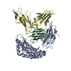 1p7qS S: Starting model for refinement |
|---|---|
| Similar structure data |


-Links
 PDBj
PDBj


- Assembly
| Deposited unit | 
| ||||||||
|---|---|---|---|---|---|---|---|---|---|
| 1 | 
| ||||||||
| 2 | 
| ||||||||
| Unit cell |
|
-Components
-Protein , 2 types, 4 molecules BFDH
| #2: Protein | Mass: 11748.160 Da / Num. of mol.: 2 / Source method: isolated from a natural source / Details: beta2m / Source: (natural) Homo sapiens (human) / References: UniProt: P61769#3: Protein | Mass: 22055.697 Da / Num. of mol.: 2 / Fragment: Ig-like C2-type 1 and C2-type 2 domains / Source method: isolated from a natural source / Details: LIR-1 / Source: (natural) Homo sapiens (human) / References: UniProt: Q8NHL6 |
|---|
-Antibody / Protein/peptide / Non-polymers , 3 types, 366 molecules AEGC
| #1: Antibody | Mass: 31664.994 Da / Num. of mol.: 2 / Fragment: sequence database residues 21-301 / Source method: isolated from a natural source / Details: gene UL18 / Source: (natural) Human herpesvirus 5 / Strain: AD169 / References: UniProt: Q4A1U8, UniProt: P08560*PLUS#4: Protein/peptide | Mass: 1005.257 Da / Num. of mol.: 2 / Fragment: sequence database residues 170-178 / Source method: obtained synthetically / Source: (synth.) Homo sapiens (human) / References: UniProt: P60709#9: Water | ChemComp-HOH / | Mass: 18.015 Da / Num. of mol.: 362 / Source method: isolated from a natural source / Formula: H2O |
|---|
-Sugars , 4 types, 16 molecules 


| #5: Sugar | ChemComp-NAG /  Type: D-saccharide, beta linking / Mass: 221.208 Da / Num. of mol.: 13 Type: D-saccharide, beta linking / Mass: 221.208 Da / Num. of mol.: 13Source method: isolated from a genetically manipulated source Formula: C8H15NO6 #6: Sugar | ChemComp-MAN / |  Type: D-saccharide, alpha linking / Mass: 180.156 Da / Num. of mol.: 1 Type: D-saccharide, alpha linking / Mass: 180.156 Da / Num. of mol.: 1Source method: isolated from a genetically manipulated source Formula: C6H12O6 #7: Sugar | ChemComp-BMA / |  Type: D-saccharide, beta linking / Mass: 180.156 Da / Num. of mol.: 1 Type: D-saccharide, beta linking / Mass: 180.156 Da / Num. of mol.: 1Source method: isolated from a genetically manipulated source Formula: C6H12O6 #8: Sugar | ChemComp-FUC / |  Type: L-saccharide, alpha linking / Mass: 164.156 Da / Num. of mol.: 1 Type: L-saccharide, alpha linking / Mass: 164.156 Da / Num. of mol.: 1Source method: isolated from a genetically manipulated source Formula: C6H12O5 |
|---|
-Details
| Has protein modification | Y |
|---|
-Experimental details
-Experiment
| Experiment | Method: X-RAY DIFFRACTION / Number of used crystals: 1 |
|---|
- Sample preparation
| Crystal | Density Matthews: 3.29 Å3/Da / Density % sol: 62.61 % |
|---|---|
| Crystal grow | Temperature: 298 K / Method: vapor diffusion, hanging drop / pH: 5.5 Details: 0.4 M Mg(NO3)2, and 16~22% (w/v) PEG 33500, pH 5.5, VAPOR DIFFUSION, HANGING DROP, temperature 298K |
-Data collection
| Diffraction | Mean temperature: 100 K |
|---|---|
| Diffraction source | Source: SYNCHROTRON / Site: SSRL  / Beamline: BL9-2 / Wavelength: 1 Å / Beamline: BL9-2 / Wavelength: 1 Å |
| Detector | Type: MARMOSAIC 325 mm CCD / Detector: CCD / Date: Mar 30, 2007 / Details: mirrors |
| Radiation | Protocol: SINGLE WAVELENGTH / Monochromatic (M) / Laue (L): M / Scattering type: x-ray |
| Radiation wavelength | Wavelength: 1 Å / Relative weight: 1 |
| Reflection | Resolution: 2.21→50 Å / Num. obs: 85873 / % possible obs: 98.6 % / Rmerge(I) obs: 0.043 |
| Reflection shell | Resolution: 2.21→2.29 Å / Redundancy: 3.5 % / Rmerge(I) obs: 0.239 / Mean I/σ(I) obs: 6.6 / Num. unique all: 7747 / % possible all: 89.2 |
-Phasing
| Phasing | Method: molecular replacement |
|---|
- Processing
| Software |
| ||||||||||||||||||||||||||||||||
|---|---|---|---|---|---|---|---|---|---|---|---|---|---|---|---|---|---|---|---|---|---|---|---|---|---|---|---|---|---|---|---|---|---|
| Refinement | Method to determine structure: MOLECULAR REPLACEMENT Starting model: PDB entry 1P7Q Resolution: 2.21→47.46 Å / Isotropic thermal model: Isotropic / Cross valid method: THROUGHOUT / σ(F): 0 / σ(I): 0 / Stereochemistry target values: Engh & Huber
| ||||||||||||||||||||||||||||||||
| Displacement parameters | Biso mean: 42.508 Å2 | ||||||||||||||||||||||||||||||||
| Refine analyze | Luzzati coordinate error obs: 0.031 Å | ||||||||||||||||||||||||||||||||
| Refinement step | Cycle: LAST / Resolution: 2.21→47.46 Å
| ||||||||||||||||||||||||||||||||
| Refine LS restraints |
|
