 Movie
Movie Controller

[English] 日本語
 Yorodumi
- PDB-3c7g: Crystal structure of a glycoside hydrolase family 43 arabinoxylan... -
+ Open data

- Basic information
| Entry | Database: PDB / ID: 3c7g | |||||||||
|---|---|---|---|---|---|---|---|---|---|---|
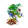
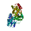
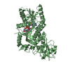
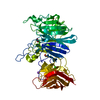
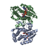
| Title | Crystal structure of a glycoside hydrolase family 43 arabinoxylan arabinofuranohydrolase from Bacillus subtilis in complex with xylotetraose. | |||||||||
 Components Components | Endo-1,4-beta-xylanase | |||||||||
 Keywords Keywords | HYDROLASE / 5-bladed beta-propeller fold / beta-sandwich / Xylan degradation | |||||||||
| Function / homology |  Function and homology information Function and homology informationnon-reducing end alpha-L-arabinofuranosidase / alpha-L-arabinofuranosidase activity / xylan catabolic process / carbohydrate binding / extracellular region / metal ion binding Similarity search - Function | |||||||||
| Biological species |  | |||||||||
| Method |  X-RAY DIFFRACTION / SYNCHROTRON / MOLECULAR REPLACEMENT / Resolution: 2.02 Å X-RAY DIFFRACTION / SYNCHROTRON / MOLECULAR REPLACEMENT / Resolution: 2.02 Å | |||||||||
 Authors Authors | Vandermarliere, E. / Bourgois, T.M. / Winn, M.D. / Van Campenhout, S. / Volckaert, G. / Strelkov, S.V. / Delcour, J.A. / Rabijns, A. / Courtin, C.M. | |||||||||
 Citation Citation | Journal: Biochem.J. / Year: 2009 Title: Structural analysis of a glycoside hydrolase family 43 arabinoxylan arabinofuranohydrolase in complex with xylotetraose reveals a different binding mechanism compared with other members of the same family. Authors: Vandermarliere, E. / Bourgois, T.M. / Winn, M.D. / van Campenhout, S. / Volckaert, G. / Delcour, J.A. / Strelkov, S.V. / Rabijns, A. / Courtin, C.M. | |||||||||
| History |
|
- Structure visualization
| Structure viewer | Molecule: MolmilJmol/JSmol |
|---|
- Downloads & links
-Download
| PDBx/mmCIF format | 3c7g.cif.gz | 119.6 KB | Display | PDBx/mmCIF format |
|---|---|---|---|---|
| PDB format | pdb3c7g.ent.gz | 88.9 KB | Display | PDB format |
| PDBx/mmJSON format | 3c7g.json.gz | Tree view | PDBx/mmJSON format | |
| Others |  Other downloads Other downloads |
-Validation report
| Arichive directory | https://data.pdbj.org/pub/pdb/validation_reports/c7/3c7gftp://data.pdbj.org/pub/pdb/validation_reports/c7/3c7g | HTTPS FTP |
|---|
-Related structure data
| Related structure data |  3c7eC  3c7fC 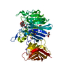 3c7hC  3c7oC  1gyhS  1w9tS C: citing same article ( S: Starting model for refinement |
|---|---|
| Similar structure data |


-Links
 PDBj
PDBj- Assembly
| Deposited unit | 
| ||||||||
|---|---|---|---|---|---|---|---|---|---|
| 1 |
| ||||||||
| Unit cell |
|
-Components
-Protein / Sugars , 2 types, 2 molecules A
| #1: Protein | Mass: 51763.164 Da / Num. of mol.: 1 Source method: isolated from a genetically manipulated source Source: (gene. exp.) References: UniProt: Q45071, non-reducing end alpha-L-arabinofuranosidase |
|---|---|
| #2: Polysaccharide | beta-D-xylopyranose-(1-4)-beta-D-xylopyranose-(1-4)-beta-D-xylopyranose-(1-4)-beta-D-xylopyranose Source method: isolated from a genetically manipulated source |
-Non-polymers , 4 types, 450 molecules 


| #3: Chemical | ChemComp-CA /  Mass: 40.078 Da / Num. of mol.: 1 / Source method: obtained synthetically / Formula: Ca Mass: 40.078 Da / Num. of mol.: 1 / Source method: obtained synthetically / Formula: Ca | ||||
|---|---|---|---|---|---|
| #4: Chemical |  Mass: 22.990 Da / Num. of mol.: 2 / Source method: obtained synthetically / Formula: Na Mass: 22.990 Da / Num. of mol.: 2 / Source method: obtained synthetically / Formula: Na#5: Chemical | ChemComp-GOL / |  Mass: 92.094 Da / Num. of mol.: 1 / Source method: obtained synthetically / Formula: C3H8O3 Mass: 92.094 Da / Num. of mol.: 1 / Source method: obtained synthetically / Formula: C3H8O3#6: Water | ChemComp-HOH / | Mass: 18.015 Da / Num. of mol.: 446 / Source method: isolated from a natural source / Formula: H2O |
-Experimental details
-Experiment
| Experiment | Method: X-RAY DIFFRACTION / Number of used crystals: 1 |
|---|
- Sample preparation
| Crystal | Density Matthews: 2.52 Å3/Da / Density % sol: 51.25 % |
|---|---|
| Crystal grow | Temperature: 277 K / Method: vapor diffusion, hanging drop / pH: 5 Details: 4.0 M Na formate , pH 5.0, VAPOR DIFFUSION, HANGING DROP, temperature 277K |
-Data collection
| Diffraction | Mean temperature: 100 K |
|---|---|
| Diffraction source | Source: SYNCHROTRON / Site: EMBL/DESY, HAMBURG  / Beamline: BW7A / Wavelength: 1.0322 Å / Beamline: BW7A / Wavelength: 1.0322 Å |
| Detector | Type: MAR CCD 165 mm / Detector: CCD |
| Radiation | Protocol: SINGLE WAVELENGTH / Monochromatic (M) / Laue (L): M / Scattering type: x-ray |
| Radiation wavelength | Wavelength: 1.0322 Å / Relative weight: 1 |
| Reflection | Resolution: 2.02→50 Å / Num. obs: 33537 / % possible obs: 97 % / Redundancy: 4.5 % / Rmerge(I) obs: 0.046 / Rsym value: 4.6 |
| Reflection shell | Resolution: 2.02→2.09 Å / Redundancy: 4.2 % / Rmerge(I) obs: 0.137 / Mean I/σ(I) obs: 8 / Num. unique all: 1596 / Rsym value: 13.7 / % possible all: 93.9 |
- Processing
| Software |
| |||||||||||||||||||||||||||||||||||||||||||||||||||||||||||||||||||||||||||||||||||||||||||||||
|---|---|---|---|---|---|---|---|---|---|---|---|---|---|---|---|---|---|---|---|---|---|---|---|---|---|---|---|---|---|---|---|---|---|---|---|---|---|---|---|---|---|---|---|---|---|---|---|---|---|---|---|---|---|---|---|---|---|---|---|---|---|---|---|---|---|---|---|---|---|---|---|---|---|---|---|---|---|---|---|---|---|---|---|---|---|---|---|---|---|---|---|---|---|---|---|---|
| Refinement | Method to determine structure: MOLECULAR REPLACEMENT Starting model: pdb entries 1gyh, 1w9t Resolution: 2.02→31.96 Å / Cor.coef. Fo:Fc: 0.962 / Cor.coef. Fo:Fc free: 0.939 / SU B: 2.862 / SU ML: 0.081 / Cross valid method: THROUGHOUT / ESU R: 0.157 / ESU R Free: 0.142 / Stereochemistry target values: MAXIMUM LIKELIHOOD / Details: HYDROGENS HAVE BEEN ADDED IN THE RIDING POSITIONS
| |||||||||||||||||||||||||||||||||||||||||||||||||||||||||||||||||||||||||||||||||||||||||||||||
| Solvent computation | Ion probe radii: 0.8 Å / Shrinkage radii: 0.8 Å / VDW probe radii: 1.2 Å / Solvent model: MASK | |||||||||||||||||||||||||||||||||||||||||||||||||||||||||||||||||||||||||||||||||||||||||||||||
| Displacement parameters | Biso mean: 13.734 Å2
| |||||||||||||||||||||||||||||||||||||||||||||||||||||||||||||||||||||||||||||||||||||||||||||||
| Refinement step | Cycle: LAST / Resolution: 2.02→31.96 Å
| |||||||||||||||||||||||||||||||||||||||||||||||||||||||||||||||||||||||||||||||||||||||||||||||
| Refine LS restraints |
| |||||||||||||||||||||||||||||||||||||||||||||||||||||||||||||||||||||||||||||||||||||||||||||||
| LS refinement shell | Resolution: 2.02→2.069 Å / Total num. of bins used: 20
|
