| 登録情報 | データベース: PDB / ID: 3bxr
|
|---|
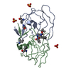
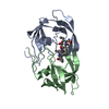
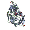
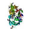
| タイトル | Crystal Structures Of Highly Constrained Substrate And Hydrolysis Products Bound To HIV-1 Protease. Implications For Catalytic Mechanism |
|---|
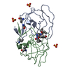
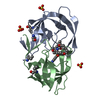
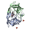
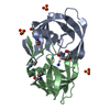
 要素 要素 | Protease |
|---|
 キーワード キーワード | HYDROLASE / HIV protease / HIVPR / substrate / product / AIDS / Aspartyl protease / Capsid maturation / Core protein / Cytoplasm / DNA integration / DNA recombination / DNA-directed DNA polymerase / Endonuclease / Lipoprotein / Magnesium / Membrane / Metal-binding / Multifunctional enzyme / Myristate / Nuclease / Nucleotidyltransferase / Nucleus / Phosphoprotein / RNA-binding / RNA-directed DNA polymerase / Transferase / Viral nucleoprotein / Virion / Zinc / Zinc-finger |
|---|
| 機能・相同性 |  機能・相同性情報 機能・相同性情報
HIV-1 retropepsin / symbiont-mediated activation of host apoptosis / retroviral ribonuclease H / exoribonuclease H / exoribonuclease H activity / host multivesicular body / DNA integration / viral genome integration into host DNA / RNA-directed DNA polymerase / establishment of integrated proviral latency ...HIV-1 retropepsin / symbiont-mediated activation of host apoptosis / retroviral ribonuclease H / exoribonuclease H / exoribonuclease H activity / host multivesicular body / DNA integration / viral genome integration into host DNA / RNA-directed DNA polymerase / establishment of integrated proviral latency / viral penetration into host nucleus / RNA stem-loop binding / RNA-directed DNA polymerase activity / RNA-DNA hybrid ribonuclease activity / 転移酵素; リンを含む基を移すもの; 核酸を移すもの / host cell / viral nucleocapsid / DNA recombination / DNA-directed DNA polymerase / aspartic-type endopeptidase activity / 加水分解酵素; エステル加水分解酵素 / DNA-directed DNA polymerase activity / symbiont-mediated suppression of host gene expression / viral translational frameshifting / lipid binding / symbiont entry into host cell / host cell nucleus / host cell plasma membrane / virion membrane / structural molecule activity / proteolysis / DNA binding / zinc ion binding / membrane類似検索 - 分子機能 Reverse transcriptase connection / Reverse transcriptase connection domain / Reverse transcriptase thumb / Reverse transcriptase thumb domain / Integrase Zinc binding domain / Zinc finger integrase-type profile. / Integrase-like, N-terminal / Integrase DNA binding domain / Integrase, C-terminal domain superfamily, retroviral / Integrase, N-terminal zinc-binding domain ...Reverse transcriptase connection / Reverse transcriptase connection domain / Reverse transcriptase thumb / Reverse transcriptase thumb domain / Integrase Zinc binding domain / Zinc finger integrase-type profile. / Integrase-like, N-terminal / Integrase DNA binding domain / Integrase, C-terminal domain superfamily, retroviral / Integrase, N-terminal zinc-binding domain / Integrase, C-terminal, retroviral / Integrase DNA binding domain profile. / Immunodeficiency lentiviral matrix, N-terminal / gag gene protein p17 (matrix protein) / RNase H / Integrase core domain / Integrase, catalytic core / Integrase catalytic domain profile. / Retropepsin-like catalytic domain / Matrix protein, lentiviral and alpha-retroviral, N-terminal / Retroviral nucleocapsid Gag protein p24, C-terminal domain / Gag protein p24 C-terminal domain / RNase H type-1 domain profile. / Ribonuclease H domain / Retropepsins / Retroviral aspartyl protease / Aspartyl protease, retroviral-type family profile. / Peptidase A2A, retrovirus, catalytic / Reverse transcriptase domain / Reverse transcriptase (RNA-dependent DNA polymerase) / Reverse transcriptase (RT) catalytic domain profile. / Retrovirus capsid, C-terminal / Retroviral matrix protein / Cathepsin D, subunit A; domain 1 / Acid Proteases / Retrovirus capsid, N-terminal / zinc finger / Zinc knuckle / Zinc finger, CCHC-type superfamily / Zinc finger, CCHC-type / Zinc finger CCHC-type profile. / Aspartic peptidase, active site / Eukaryotic and viral aspartyl proteases active site. / Aspartic peptidase domain superfamily / Ribonuclease H superfamily / Ribonuclease H-like superfamily / Reverse transcriptase/Diguanylate cyclase domain / DNA/RNA polymerase superfamily / Beta Barrel / Mainly Beta類似検索 - ドメイン・相同性 |
|---|
| 手法 |  X線回折 / フーリエ合成 / 解像度: 1.6 Å X線回折 / フーリエ合成 / 解像度: 1.6 Å |
|---|
 データ登録者 データ登録者 | Tyndall, J.D. / Pattenden, L.K. / Reid, R.C. / Hu, S.H. / Alewood, D. / Alewood, P.F. / Walsh, T. / Fairlie, D.P. / Martin, J.L. |
|---|
 引用 引用 | #1: ジャーナル: Biochemistry / 年: 1999タイトル: Molecular recognition of macrocyclic peptidomimetic inhibitors by HIV-1 protease 著者: Martin, J.L. / Begun, J. / Schindeler, A. / Wickramasinghe, W.A. / Alewood, D. / Alewood, P.F. / Bergman, D.A. / Brinkworth, R.I. / Abbenante, G. / March, D.R. / Reid, R.C. / Fairlie, D.P. |
|---|
| 履歴 | | 登録 | 2008年1月14日 | 登録サイト: RCSB / 処理サイト: PDBJ |
|---|
| 改定 1.0 | 2008年3月25日 | Provider: repository / タイプ: Initial release |
|---|
| 改定 1.1 | 2011年7月13日 | Group: Version format compliance |
|---|
| 改定 1.2 | 2017年10月25日 | Group: Refinement description / カテゴリ: software |
|---|
| 改定 1.3 | 2021年11月10日 | Group: Database references / Derived calculations
カテゴリ: database_2 / struct_conn ...database_2 / struct_conn / struct_ref_seq_dif / struct_site
Item: _database_2.pdbx_DOI / _database_2.pdbx_database_accession ..._database_2.pdbx_DOI / _database_2.pdbx_database_accession / _struct_conn.pdbx_leaving_atom_flag / _struct_ref_seq_dif.details / _struct_site.pdbx_auth_asym_id / _struct_site.pdbx_auth_comp_id / _struct_site.pdbx_auth_seq_id |
|---|
| 改定 1.4 | 2023年11月1日 | Group: Data collection / Refinement description
カテゴリ: chem_comp_atom / chem_comp_bond / pdbx_initial_refinement_model |
|---|
| 改定 1.5 | 2023年11月15日 | Group: Data collection / カテゴリ: chem_comp_atom / chem_comp_bond / Item: _chem_comp_atom.atom_id / _chem_comp_bond.atom_id_2 |
|---|
|
|---|
 ムービー
ムービー コントローラー


 データを開く
 基本情報
基本情報 構造の表示
構造の表示 ダウンロードとリンク
 その他のダウンロード


 PDBj
PDBj

 集合体
集合体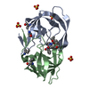

 分子量: 96.063 Da / 分子数: 4 / 由来タイプ: 合成 / 式: SO4
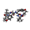
 分子量: 677.830 Da / 分子数: 1 / 由来タイプ: 合成 / 式: C37H51N5O7
 分子量: 18.015 Da / 分子数: 207 / 由来タイプ: 天然 / 式: H2O
 試料調製
試料調製 解析
解析