 Movie
Movie Controller

[English] 日本語
 Yorodumi
- PDB-3bam: RESTRICTION ENDONUCLEASE BAMHI COMPLEX WITH DNA AND MANGANESE ION... -
+ Open data

- Basic information
| Entry | Database: PDB / ID: 3bam | ||||||
|---|---|---|---|---|---|---|---|
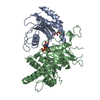
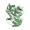
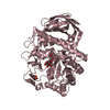
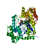
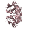
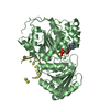
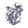
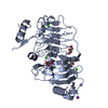
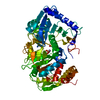
| Title | RESTRICTION ENDONUCLEASE BAMHI COMPLEX WITH DNA AND MANGANESE IONS (POST-REACTIVE COMPLEX) | ||||||
 Components Components |
| ||||||
 Keywords Keywords | HYDROLASE/DNA / HYDROLASE / PHOSPHODIESTERASE / COMPLEX (ENDONUCLEASE-DNA) / NUCLEASE / PROTEIN/DNA / HYDROLASE-DNA COMPLEX | ||||||
| Function / homology |  Function and homology information Function and homology informationtype II site-specific deoxyribonuclease / type II site-specific deoxyribonuclease activity / DNA restriction-modification system / magnesium ion binding / DNA binding Similarity search - Function | ||||||
| Biological species |  | ||||||
| Method |  X-RAY DIFFRACTION / SYNCHROTRON / MOLECULAR REPLACEMENT / Resolution: 1.8 Å X-RAY DIFFRACTION / SYNCHROTRON / MOLECULAR REPLACEMENT / Resolution: 1.8 Å | ||||||
 Authors Authors | Viadiu, H. / Aggarwal, A.K. | ||||||
 Citation Citation | Journal: Nat.Struct.Biol. / Year: 1998 Title: The role of metals in catalysis by the restriction endonuclease BamHI. Authors: Viadiu, H. / Aggarwal, A.K. #1: Journal: Science / Year: 1995Title: Structure of Bam Hi Endonuclease Bound to DNA: Partial Folding and Unfolding on DNA Binding Authors: Newman, M. / Strzelecka, T. / Dorner, L.F. / Schildkraut, I. / Aggarwal, A.K. | ||||||
| History |
|
- Structure visualization
| Structure viewer | Molecule: MolmilJmol/JSmol |
|---|
- Downloads & links
-Download
| PDBx/mmCIF format | 3bam.cif.gz | 117.9 KB | Display | PDBx/mmCIF format |
|---|---|---|---|---|
| PDB format | pdb3bam.ent.gz | 88.2 KB | Display | PDB format |
| PDBx/mmJSON format | 3bam.json.gz | Tree view | PDBx/mmJSON format | |
| Others |  Other downloads Other downloads |
-Validation report
| Arichive directory | https://data.pdbj.org/pub/pdb/validation_reports/ba/3bamftp://data.pdbj.org/pub/pdb/validation_reports/ba/3bam | HTTPS FTP |
|---|
-Related structure data
| Related structure data | 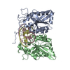 2bamC  1bhmS S: Starting model for refinement C: citing same article ( |
|---|---|
| Similar structure data |

-Links
 PDBj
PDBj


- Assembly
| Deposited unit | 
| ||||||||
|---|---|---|---|---|---|---|---|---|---|
| 1 |
| ||||||||
| Unit cell |
| ||||||||
| Details | THE CRYSTALLOGRAPHIC ASYMMETRIC UNIT CONTAINS TWO BAMHI MONOMERS (CHAINS A AND B) BOUND TO A PALINDROMIC 12 BASE PAIR DNA COMPLEX. |
-Components
-DNA chain , 3 types, 3 molecules CDE
| #1: DNA chain | Mass: 3661.416 Da / Num. of mol.: 1 / Source method: obtained synthetically |
|---|---|
| #2: DNA chain | Mass: 1205.841 Da / Num. of mol.: 1 / Source method: obtained synthetically |
| #3: DNA chain | Mass: 2410.618 Da / Num. of mol.: 1 / Source method: obtained synthetically |
-Protein , 1 types, 2 molecules AB
| #4: Protein | Mass: 24602.297 Da / Num. of mol.: 2 Source method: isolated from a genetically manipulated source Details: CRYSTAL GROWN IN THE PRESENCE OF MANGANESE IONS. / Source: (gene. exp.) References: UniProt: P23940, type II site-specific deoxyribonuclease |
|---|
-Non-polymers , 2 types, 354 molecules 

| #5: Chemical |  Mass: 54.938 Da / Num. of mol.: 2 / Source method: obtained synthetically / Formula: Mn Mass: 54.938 Da / Num. of mol.: 2 / Source method: obtained synthetically / Formula: Mn#6: Water | ChemComp-HOH / | Mass: 18.015 Da / Num. of mol.: 352 / Source method: isolated from a natural source / Formula: H2O |
|---|
-Details
| Compound details | THE C-TERMINALS ARE ASYMMETRIC |
|---|
-Experimental details
-Experiment
| Experiment | Method: X-RAY DIFFRACTION / Number of used crystals: 1 |
|---|
- Sample preparation
| Crystal | Density Matthews: 2.52 Å3/Da / Density % sol: 51.22 % / Description: CRYSTAL WAS ISOMORPHOUS TO 1BHM. | |||||||||||||||||||||||||||||||||||||||||||||||||||||||||||||||||||||||||||||||||||||||||||
|---|---|---|---|---|---|---|---|---|---|---|---|---|---|---|---|---|---|---|---|---|---|---|---|---|---|---|---|---|---|---|---|---|---|---|---|---|---|---|---|---|---|---|---|---|---|---|---|---|---|---|---|---|---|---|---|---|---|---|---|---|---|---|---|---|---|---|---|---|---|---|---|---|---|---|---|---|---|---|---|---|---|---|---|---|---|---|---|---|---|---|---|---|
| Crystal grow | pH: 5.3 Details: pH 5.3 CATALYSIS OCCURRED IN THE CRYSTAL AFTER SOAKING MANGANESE ION. THE ACTIVE SITE OF THE R SUBUNIT CLEAVED THE ORIGINAL D STRAND IN TWO (D AND E) RESULTING IN A POST-REACTIVE COMPLEX. | |||||||||||||||||||||||||||||||||||||||||||||||||||||||||||||||||||||||||||||||||||||||||||
| Crystal grow | *PLUS Temperature: 20 ℃ / Method: vapor diffusion, hanging drop | |||||||||||||||||||||||||||||||||||||||||||||||||||||||||||||||||||||||||||||||||||||||||||
| Components of the solutions | *PLUS
|
-Data collection
| Diffraction | Mean temperature: 110 K |
|---|---|
| Diffraction source | Source: SYNCHROTRON / Site: CHESS  / Beamline: F1 / Wavelength: 0.908 / Beamline: F1 / Wavelength: 0.908 |
| Detector | Detector: IMAGE PLATE |
| Radiation | Protocol: SINGLE WAVELENGTH / Monochromatic (M) / Laue (L): M / Scattering type: x-ray |
| Radiation wavelength | Wavelength: 0.908 Å / Relative weight: 1 |
| Reflection | Resolution: 1.8→40 Å / Num. obs: 40212 / % possible obs: 72 % / Observed criterion σ(I): 0 / Redundancy: 2.77 % / Rmerge(I) obs: 0.084 / Rsym value: 0.084 / Net I/σ(I): 8.09 |
| Reflection | *PLUS Num. measured all: 111430 |
| Reflection shell | *PLUS % possible obs: 45.6 % / Rmerge(I) obs: 0.318 |
- Processing
| Software |
| ||||||||||||||||||||||||||||||||||||||||||||||||||||||||||||
|---|---|---|---|---|---|---|---|---|---|---|---|---|---|---|---|---|---|---|---|---|---|---|---|---|---|---|---|---|---|---|---|---|---|---|---|---|---|---|---|---|---|---|---|---|---|---|---|---|---|---|---|---|---|---|---|---|---|---|---|---|---|
| Refinement | Method to determine structure: MOLECULAR REPLACEMENT Starting model: 1BHM Resolution: 1.8→8 Å / Rfactor Rfree error: 0 / Cross valid method: R-FREE / σ(F): 2 Details: THE MANGANESE METALS WERE BOUND ONLY IN ONE ACTIVE SITE OF THE DIMER (THE R-SUBUNIT).
| ||||||||||||||||||||||||||||||||||||||||||||||||||||||||||||
| Displacement parameters | Biso mean: 30 Å2 | ||||||||||||||||||||||||||||||||||||||||||||||||||||||||||||
| Refinement step | Cycle: LAST / Resolution: 1.8→8 Å
| ||||||||||||||||||||||||||||||||||||||||||||||||||||||||||||
| Refine LS restraints |
| ||||||||||||||||||||||||||||||||||||||||||||||||||||||||||||
| Xplor file |
| ||||||||||||||||||||||||||||||||||||||||||||||||||||||||||||
| Software | *PLUS Name: X-PLOR / Version: 3.851 / Classification: refinement | ||||||||||||||||||||||||||||||||||||||||||||||||||||||||||||
| Refinement | *PLUS Highest resolution: 1.8 Å / Lowest resolution: 8 Å / σ(F): 2 / % reflection Rfree: 3 % | ||||||||||||||||||||||||||||||||||||||||||||||||||||||||||||
| Solvent computation | *PLUS | ||||||||||||||||||||||||||||||||||||||||||||||||||||||||||||
| Displacement parameters | *PLUS Biso mean: 30 Å2 | ||||||||||||||||||||||||||||||||||||||||||||||||||||||||||||
| Refine LS restraints | *PLUS
|
