 Movie
Movie Controller

[English] 日本語
 Yorodumi
- PDB-1mf1: Structure of the Recombinant Mouse-Muscle Adenylosuccinate Synthe... -
+ Open data

- Basic information
| Entry | Database: PDB / ID: 1mf1 | ||||||
|---|---|---|---|---|---|---|---|
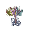
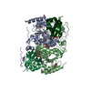
| Title | Structure of the Recombinant Mouse-Muscle Adenylosuccinate Synthetase Complexed with AMP | ||||||
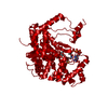
 Components Components | Adenylosuccinate Synthetase | ||||||
 Keywords Keywords | LIGASE / Purine biosynthesis / GTP-binding / Multigene family | ||||||
| Function / homology |  Function and homology information Function and homology informationPurine ribonucleoside monophosphate biosynthesis / AMP biosynthetic process / adenylosuccinate synthase / adenylosuccinate synthase activity / purine nucleotide metabolic process / aspartate metabolic process / cellular response to electrical stimulus / IMP metabolic process / 'de novo' AMP biosynthetic process / AMP salvage ...Purine ribonucleoside monophosphate biosynthesis / AMP biosynthetic process / adenylosuccinate synthase / adenylosuccinate synthase activity / purine nucleotide metabolic process / aspartate metabolic process / cellular response to electrical stimulus / IMP metabolic process / 'de novo' AMP biosynthetic process / AMP salvage / L-glutamine metabolic process / response to starvation / response to muscle activity / cellular response to xenobiotic stimulus / actin filament binding / GTPase activity / GTP binding / magnesium ion binding / membrane / identical protein binding / cytoplasm Similarity search - Function | ||||||
| Biological species |  | ||||||
| Method |  X-RAY DIFFRACTION / MOLECULAR REPLACEMENT / Resolution: 2.7 Å X-RAY DIFFRACTION / MOLECULAR REPLACEMENT / Resolution: 2.7 Å | ||||||
 Authors Authors | Iancu, C.V. / Borza, T. / Fromm, H.J. / Honzatko, R.B. | ||||||
 Citation Citation | Journal: J.Biol.Chem. / Year: 2002 Title: Feedback inhibition and product complexes of recombinant mouse muscle adenylosuccinate synthetase. Authors: Iancu, C.V. / Borza, T. / Fromm, H.J. / Honzatko, R.B. | ||||||
| History |
|
- Structure visualization
| Structure viewer | Molecule: MolmilJmol/JSmol |
|---|
- Downloads & links
-Download
| PDBx/mmCIF format | 1mf1.cif.gz | 95.9 KB | Display | PDBx/mmCIF format |
|---|---|---|---|---|
| PDB format | pdb1mf1.ent.gz | 73.6 KB | Display | PDB format |
| PDBx/mmJSON format | 1mf1.json.gz | Tree view | PDBx/mmJSON format | |
| Others |  Other downloads Other downloads |
-Validation report
| Arichive directory | https://data.pdbj.org/pub/pdb/validation_reports/mf/1mf1ftp://data.pdbj.org/pub/pdb/validation_reports/mf/1mf1 | HTTPS FTP |
|---|
-Related structure data


-Links
 PDBj
PDBj


- Assembly
| Deposited unit | 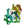
| ||||||||
|---|---|---|---|---|---|---|---|---|---|
| 1 | 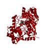
| ||||||||
| Unit cell |
| ||||||||
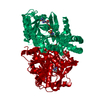
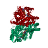
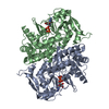
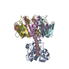
| Details | The biological unit is a dimer. The other monomer is generated by:2-y, 2-x, 1/2-z. |
-Components
| #1: Protein | Mass: 50321.301 Da / Num. of mol.: 1 Source method: isolated from a genetically manipulated source Source: (gene. exp.)  |
|---|---|
| #2: Chemical | ChemComp-ACT /   Mass: 59.044 Da / Num. of mol.: 1 / Source method: obtained synthetically / Formula: C2H3O2 Mass: 59.044 Da / Num. of mol.: 1 / Source method: obtained synthetically / Formula: C2H3O2 |
| #3: Chemical | ChemComp-AMP /   Mass: 347.221 Da / Num. of mol.: 1 / Source method: obtained synthetically / Formula: C10H14N5O7P / Comment: AMP*YM Mass: 347.221 Da / Num. of mol.: 1 / Source method: obtained synthetically / Formula: C10H14N5O7P / Comment: AMP*YM |
| #4: Water | ChemComp-HOH /  Mass: 18.015 Da / Num. of mol.: 45 / Source method: isolated from a natural source / Formula: H2O Mass: 18.015 Da / Num. of mol.: 45 / Source method: isolated from a natural source / Formula: H2O |
-Experimental details
-Experiment
| Experiment | Method: X-RAY DIFFRACTION / Number of used crystals: 1 |
|---|
- Sample preparation
| Crystal | Density Matthews: 2.44 Å3/Da / Density % sol: 49.58 % | |||||||||||||||||||||||||||||||||||||||||||||||||||||||||||||||||||||||||||||
|---|---|---|---|---|---|---|---|---|---|---|---|---|---|---|---|---|---|---|---|---|---|---|---|---|---|---|---|---|---|---|---|---|---|---|---|---|---|---|---|---|---|---|---|---|---|---|---|---|---|---|---|---|---|---|---|---|---|---|---|---|---|---|---|---|---|---|---|---|---|---|---|---|---|---|---|---|---|---|
| Crystal grow | Temperature: 298 K / Method: vapor diffusion, hanging drop / pH: 7 Details: PEG, pH 7, VAPOR DIFFUSION, HANGING DROP, temperature 298K | |||||||||||||||||||||||||||||||||||||||||||||||||||||||||||||||||||||||||||||
| Crystal grow | *PLUS | |||||||||||||||||||||||||||||||||||||||||||||||||||||||||||||||||||||||||||||
| Components of the solutions | *PLUS
|
-Data collection
| Diffraction | Mean temperature: 100 K |
|---|---|
| Diffraction source | Source: ROTATING ANODE / Type: RIGAKU |
| Detector | Type: RIGAKU / Detector: IMAGE PLATE |
| Radiation | Protocol: SINGLE WAVELENGTH / Monochromatic (M) / Laue (L): M / Scattering type: x-ray |
| Radiation wavelength | Relative weight: 1 |
| Reflection | Resolution: 2.7→49.7 Å / Num. all: 14816 / Num. obs: 14742 / % possible obs: 99.5 % / Observed criterion σ(F): 2 / Observed criterion σ(I): 2 / Redundancy: 10 % / Rmerge(I) obs: 0.06 / Net I/σ(I): 7.5 |
| Reflection shell | Resolution: 2.7→2.8 Å / % possible all: 99.9 |
| Reflection | *PLUS Num. measured all: 152538 / Rmerge(I) obs: 0.062 |
| Reflection shell | *PLUS % possible obs: 99.9 % / Rmerge(I) obs: 0.33 |
- Processing
| Software |
| ||||||||||||||||||||
|---|---|---|---|---|---|---|---|---|---|---|---|---|---|---|---|---|---|---|---|---|---|
| Refinement | Method to determine structure: MOLECULAR REPLACEMENT / Resolution: 2.7→15 Å / σ(F): 2 / Stereochemistry target values: Engh & Huber
| ||||||||||||||||||||
| Refinement step | Cycle: LAST / Resolution: 2.7→15 Å
| ||||||||||||||||||||
| Refine LS restraints |
| ||||||||||||||||||||
| LS refinement shell | Resolution: 2.7→2.87 Å / Rfactor Rfree error: 0.027
| ||||||||||||||||||||
| Refinement | *PLUS Lowest resolution: 15 Å / % reflection Rfree: 10 % / Rfactor Rfree: 0.265 / Rfactor Rwork: 0.219 | ||||||||||||||||||||
| Solvent computation | *PLUS | ||||||||||||||||||||
| Displacement parameters | *PLUS | ||||||||||||||||||||
| Refine LS restraints | *PLUS
| ||||||||||||||||||||
| LS refinement shell | *PLUS Rfactor Rfree: 0.397 / Rfactor Rwork: 0.345 |
