 Movie
Movie Controller

[English] 日本語
 Yorodumi
- PDB-1ltb: 2.6 ANGSTROMS CRYSTAL STRUCTURE OF PARTIALLY-ACTIVATED E. COLI HE... -
+ Open data

- Basic information
| Entry | Database: PDB / ID: 1ltb | ||||||
|---|---|---|---|---|---|---|---|
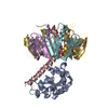
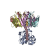
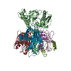
| Title | 2.6 ANGSTROMS CRYSTAL STRUCTURE OF PARTIALLY-ACTIVATED E. COLI HEAT-LABILE ENTEROTOXIN (LT) | ||||||
 Components Components |
| ||||||
 Keywords Keywords | ENTEROTOXIN | ||||||
| Function / homology |  Function and homology information Function and homology informationSecretion of toxins / toxin activity / killing of cells of another organism / : / extracellular region Similarity search - Function | ||||||
| Biological species |  | ||||||
| Method |  X-RAY DIFFRACTION / Resolution: 2.6 Å X-RAY DIFFRACTION / Resolution: 2.6 Å | ||||||
 Authors Authors | Merritt, E.A. / Sixma, T.K. / Hol, W.G.J. | ||||||
 Citation Citation | Journal: FEBS Lett. / Year: 1994 Title: Structure of partially-activated E. coli heat-labile enterotoxin (LT) at 2.6 A resolution. Authors: Merritt, E.A. / Pronk, S.E. / Sixma, T.K. / Kalk, K.H. / van Zanten, B.A. / Hol, W.G. #1: Journal: J.Mol.Biol. / Year: 1993Title: Refined Structure of Escherichia Coli Heat-Labile Enterotoxin, a Close Relative of Cholera Toxin Authors: Sixma, T.K. / Kalk, K.H. / Van Zanten, B.A.M. / Dauter, Z. / Kingma, J. / Witholt, B. / Hol, W.G.J. #2: Journal: Nature / Year: 1991Title: Crystal Structure of a Cholera Toxin-Related Heat-Labile Enterotoxin from E. Coli Authors: Sixma, T.K. / Pronk, S.E. / Kalk, K.H. / Wartna, E.S. / Van Zanten, B.A.M. / Witholt, B. / Hol, W.G.J. | ||||||
| History |
| ||||||
| Remark 700 | SHEET IN THE PENTAMER THE BETA SHEETS FROM ADJACENT MONOMERS COMBINE TO FORM A CONTINUOUS SIX- ...SHEET IN THE PENTAMER THE BETA SHEETS FROM ADJACENT MONOMERS COMBINE TO FORM A CONTINUOUS SIX-STRANDED ANTI-PARALLEL SHEET ACROSS EACH MONOMER-MONOMER INTERFACE. |
- Structure visualization
| Structure viewer | Molecule: MolmilJmol/JSmol |
|---|
- Downloads & links
-Download
| PDBx/mmCIF format | 1ltb.cif.gz | 159.3 KB | Display | PDBx/mmCIF format |
|---|---|---|---|---|
| PDB format | pdb1ltb.ent.gz | 127.3 KB | Display | PDB format |
| PDBx/mmJSON format | 1ltb.json.gz | Tree view | PDBx/mmJSON format | |
| Others |  Other downloads Other downloads |
-Validation report
| Arichive directory | https://data.pdbj.org/pub/pdb/validation_reports/lt/1ltbftp://data.pdbj.org/pub/pdb/validation_reports/lt/1ltb | HTTPS FTP |
|---|
-Related structure data
| Similar structure data |
|---|


-Links
 PDBj
PDBj
- Assembly
| Deposited unit | 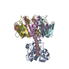
| ||||||||
|---|---|---|---|---|---|---|---|---|---|
| 1 |
| ||||||||
| Unit cell |
| ||||||||
| Atom site foot note | 1: CIS PROLINE - PRO D 93 / 2: CIS PROLINE - PRO E 93 / 3: CIS PROLINE - PRO F 93 / 4: CIS PROLINE - PRO G 93 / 5: CIS PROLINE - PRO H 93 / 6: CIS PROLINE - PRO A 178 |
-Components
| #1: Protein | Mass: 11807.539 Da / Num. of mol.: 5 Source method: isolated from a genetically manipulated source Source: (gene. exp.) #2: Protein | | Mass: 21354.600 Da / Num. of mol.: 1 Source method: isolated from a genetically manipulated source Source: (gene. exp.) #3: Protein/peptide | | Mass: 5425.951 Da / Num. of mol.: 1 Source method: isolated from a genetically manipulated source Source: (gene. exp.) #4: Water | ChemComp-HOH / |  Mass: 18.015 Da / Num. of mol.: 197 / Source method: isolated from a natural source / Formula: H2O Mass: 18.015 Da / Num. of mol.: 197 / Source method: isolated from a natural source / Formula: H2OCompound details | THIS IS THE NICKED, UNREDUCED, FORM OF THE TOXIN. THE A AND C CHAINS ARE LINKED BY A DISULFIDE ...THIS IS THE NICKED, UNREDUCED, FORM OF THE TOXIN. THE A AND C CHAINS ARE LINKED BY A DISULFIDE BRIDGE BETWEEN A 187 AND C 199, BUT THE MAIN CHAIN BETWEEN THEM HAS BEEN TRYPSIN-CLEAVED. THE MODEL IS INCOMPLETE | Has protein modification | Y | |
|---|
-Experimental details
-Experiment
| Experiment | Method: X-RAY DIFFRACTION |
|---|
- Sample preparation
| Crystal | Density Matthews: 2.21 Å3/Da / Density % sol: 44.32 % | ||||||||||||||||||||||||||||||||||||||||||||||||||||||||||||
|---|---|---|---|---|---|---|---|---|---|---|---|---|---|---|---|---|---|---|---|---|---|---|---|---|---|---|---|---|---|---|---|---|---|---|---|---|---|---|---|---|---|---|---|---|---|---|---|---|---|---|---|---|---|---|---|---|---|---|---|---|---|
| Crystal grow | *PLUS pH: 7.5 / Method: microdialysisDetails: two-layer method, taken from Pronk, S.E. et al (1985). J. Biol. Chem., 260, 13580-13584. | ||||||||||||||||||||||||||||||||||||||||||||||||||||||||||||
| Components of the solutions | *PLUS
|
-Data collection
| Radiation | Scattering type: x-ray |
|---|---|
| Radiation wavelength | Relative weight: 1 |
| Reflection | *PLUS Highest resolution: 2.6 Å / Num. obs: 21707 / % possible obs: 90 % / Num. measured all: 47662 / Rmerge(I) obs: 0.086 |
- Processing
| Software |
| ||||||||||||||||||||||||||||||||||||||||||||||||||||||||||||
|---|---|---|---|---|---|---|---|---|---|---|---|---|---|---|---|---|---|---|---|---|---|---|---|---|---|---|---|---|---|---|---|---|---|---|---|---|---|---|---|---|---|---|---|---|---|---|---|---|---|---|---|---|---|---|---|---|---|---|---|---|---|
| Refinement | Rfactor Rwork: 0.172 / Rfactor obs: 0.172 / Highest resolution: 2.6 Å | ||||||||||||||||||||||||||||||||||||||||||||||||||||||||||||
| Refinement step | Cycle: LAST / Highest resolution: 2.6 Å
| ||||||||||||||||||||||||||||||||||||||||||||||||||||||||||||
| Refine LS restraints |
| ||||||||||||||||||||||||||||||||||||||||||||||||||||||||||||
| Software | *PLUS Name: X-PLOR / Classification: refinement | ||||||||||||||||||||||||||||||||||||||||||||||||||||||||||||
| Refinement | *PLUS Lowest resolution: 10 Å / Num. reflection all: 21320 / σ(I): 0 / Rfactor all: 0.172 / Rfactor Rwork: 0.172 | ||||||||||||||||||||||||||||||||||||||||||||||||||||||||||||
| Solvent computation | *PLUS | ||||||||||||||||||||||||||||||||||||||||||||||||||||||||||||
| Displacement parameters | *PLUS | ||||||||||||||||||||||||||||||||||||||||||||||||||||||||||||
| Refine LS restraints | *PLUS Type: x_angle_d / Dev ideal: 3.24 |
