 Movie
Movie Controller

[English] 日本語
 Yorodumi
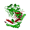
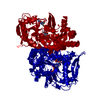
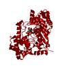
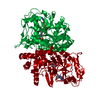
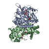
- PDB-1dj2: STRUCTURES OF ADENYLOSUCCINATE SYNTHETASE FROM TRITICUM AESTIVUM ... -
+ Open data

- Basic information
| Entry | Database: PDB / ID: 1dj2 | ||||||
|---|---|---|---|---|---|---|---|
| Title | STRUCTURES OF ADENYLOSUCCINATE SYNTHETASE FROM TRITICUM AESTIVUM AND ARABIDOPSIS THALIANA | ||||||
 Components Components | ADENYLOSUCCINATE SYNTHETASE | ||||||
 Keywords Keywords | LIGASE / GDP | ||||||
| Function / homology |  Function and homology information Function and homology informationadenylosuccinate synthase / adenylosuccinate synthase activity / IMP metabolic process / 'de novo' AMP biosynthetic process / apoplast / chloroplast stroma / plastid / chloroplast / GTP binding / magnesium ion binding / cytoplasm Similarity search - Function | ||||||
| Biological species |  | ||||||
| Method |  X-RAY DIFFRACTION / SYNCHROTRON / Resolution: 2.9 Å X-RAY DIFFRACTION / SYNCHROTRON / Resolution: 2.9 Å | ||||||
 Authors Authors | Prade, L. / Cowan-Jacob, S.W. / Chemla, P. / Potter, S. / Ward, E. / Fonne-Pfister, R. | ||||||
 Citation Citation | Journal: J.Mol.Biol. / Year: 2000 Title: Structures of adenylosuccinate synthetase from Triticum aestivum and Arabidopsis thaliana. Authors: Prade, L. / Cowan-Jacob, S.W. / Chemla, P. / Potter, S. / Ward, E. / Fonne-Pfister, R. | ||||||
| History |
|
- Structure visualization
| Structure viewer | Molecule: MolmilJmol/JSmol |
|---|
- Downloads & links
-Download
| PDBx/mmCIF format | 1dj2.cif.gz | 169.6 KB | Display | PDBx/mmCIF format |
|---|---|---|---|---|
| PDB format | pdb1dj2.ent.gz | 136.2 KB | Display | PDB format |
| PDBx/mmJSON format | 1dj2.json.gz | Tree view | PDBx/mmJSON format | |
| Others |  Other downloads Other downloads |
-Validation report
| Arichive directory | https://data.pdbj.org/pub/pdb/validation_reports/dj/1dj2ftp://data.pdbj.org/pub/pdb/validation_reports/dj/1dj2 | HTTPS FTP |
|---|
-Related structure data


-Links
 PDBj
PDBj- Assembly
| Deposited unit | 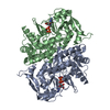
| ||||||||
|---|---|---|---|---|---|---|---|---|---|
| 1 |
| ||||||||
| Unit cell |
|
-Components
| #1: Protein | Mass: 47886.250 Da / Num. of mol.: 2 Source method: isolated from a genetically manipulated source Source: (gene. exp.)  #2: Chemical |   Type: RNA linking / Mass: 443.201 Da / Num. of mol.: 2 / Source method: obtained synthetically / Formula: C10H15N5O11P2 / Comment: GDP, energy-carrying molecule*YM Type: RNA linking / Mass: 443.201 Da / Num. of mol.: 2 / Source method: obtained synthetically / Formula: C10H15N5O11P2 / Comment: GDP, energy-carrying molecule*YM |
|---|
-Experimental details
-Experiment
| Experiment | Method: X-RAY DIFFRACTION / Number of used crystals: 1 |
|---|
- Sample preparation
| Crystal | Density Matthews: 4.19 Å3/Da / Density % sol: 70.63 % | ||||||||||||||||||||||||||||||||||||
|---|---|---|---|---|---|---|---|---|---|---|---|---|---|---|---|---|---|---|---|---|---|---|---|---|---|---|---|---|---|---|---|---|---|---|---|---|---|
| Crystal grow | Temperature: 298 K / Method: vapor diffusion, hanging drop / pH: 4.5 Details: PEG 4000, Lithium sulfate, citrate, pH 4.5, VAPOR DIFFUSION, HANGING DROP, temperature 298.0K | ||||||||||||||||||||||||||||||||||||
| Crystal grow | *PLUS pH: 7 | ||||||||||||||||||||||||||||||||||||
| Components of the solutions | *PLUS
|
-Data collection
| Diffraction | Mean temperature: 113 K |
|---|---|
| Diffraction source | Source: SYNCHROTRON / Site: ESRF  / Beamline: BM1A / Wavelength: 1 / Beamline: BM1A / Wavelength: 1 |
| Detector | Type: MARRESEARCH / Detector: IMAGE PLATE / Date: Feb 28, 1998 |
| Radiation | Protocol: SINGLE WAVELENGTH / Monochromatic (M) / Laue (L): M / Scattering type: x-ray |
| Radiation wavelength | Wavelength: 1 Å / Relative weight: 1 |
| Reflection | Resolution: 2.9→45 Å / Num. all: 33374 / Num. obs: 33374 / % possible obs: 93.8 % / Observed criterion σ(F): 2 / Observed criterion σ(I): 2 / Redundancy: 3.3 % / Biso Wilson estimate: 14.7 Å2 / Rmerge(I) obs: 0.07 / Net I/σ(I): 11.4 |
| Reflection shell | Resolution: 2.9→3 Å / Redundancy: 3.3 % / Rmerge(I) obs: 0.433 / % possible all: 87.9 |
| Reflection | *PLUS |
- Processing
| Software |
| ||||||||||||||||||||
|---|---|---|---|---|---|---|---|---|---|---|---|---|---|---|---|---|---|---|---|---|---|
| Refinement | Resolution: 2.9→45 Å / σ(F): 2 / σ(I): 2 / Stereochemistry target values: Engh & Huber
| ||||||||||||||||||||
| Refinement step | Cycle: LAST / Resolution: 2.9→45 Å
| ||||||||||||||||||||
| Refine LS restraints |
| ||||||||||||||||||||
| Software | *PLUS Name: X-PLOR / Version: 3.851 / Classification: refinement | ||||||||||||||||||||
| Refinement | *PLUS Highest resolution: 2.9 Å / Lowest resolution: 45 Å / σ(F): 2 | ||||||||||||||||||||
| Solvent computation | *PLUS | ||||||||||||||||||||
| Displacement parameters | *PLUS |
