- PDB-2zjt: Crystal structure of dna gyrase B' domain sheds lights on the mec... -
+
Open data
ID or keywords:
Loading...
-
Basic information
Entry
Database: PDB / ID: 2zjt
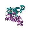
Title
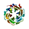
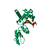
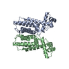
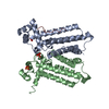
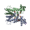
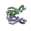
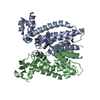
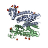
Crystal structure of dna gyrase B' domain sheds lights on the mechanism for T-segment navigation
Components
DNA gyrase subunit B
Keywords
ISOMERASE / DNA gyrase / GyrB-CTD / Toprim / Tail / DNA topoisomerase II / G-segment / T-segment / ATP-binding / Nucleotide-binding
Function / homology
Function and homology information
DNA negative supercoiling activity / DNA topoisomerase type II (double strand cut, ATP-hydrolyzing) activity / DNA topoisomerase (ATP-hydrolysing) / DNA topological change / peptidoglycan-based cell wall / DNA-templated DNA replication / chromosome / response to antibiotic / magnesium ion binding / DNA binding ...DNA negative supercoiling activity / DNA topoisomerase type II (double strand cut, ATP-hydrolyzing) activity / DNA topoisomerase (ATP-hydrolysing) / DNA topological change / peptidoglycan-based cell wall / DNA-templated DNA replication / chromosome / response to antibiotic / magnesium ion binding / DNA binding / ATP binding / metal ion binding / plasma membrane / cytoplasm Similarity search - Function
Dna Ligase; domain 1 - #440 / Rossmann fold - #670 / DNA gyrase subunit B, TOPRIM domain / DNA gyrase, subunit B / DNA topoisomerase, type IIA, subunit B / DNA gyrase B subunit, C-terminal / DNA gyrase B subunit, carboxyl terminus / DNA topoisomerase, type IIA, subunit B, domain 2 / DNA gyrase B / DNA topoisomerase, type IIA ...Dna Ligase; domain 1 - #440 / Rossmann fold - #670 / DNA gyrase subunit B, TOPRIM domain / DNA gyrase, subunit B / DNA topoisomerase, type IIA, subunit B / DNA gyrase B subunit, C-terminal / DNA gyrase B subunit, carboxyl terminus / DNA topoisomerase, type IIA, subunit B, domain 2 / DNA gyrase B / DNA topoisomerase, type IIA / DNA topoisomerase, type IIA, conserved site / DNA topoisomerase II signature. / TopoisomeraseII / DNA topoisomerase, type IIA, subunit B, C-terminal / Toprim domain / DNA topoisomerase, type IIA-like domain superfamily / Toprim domain profile. / TOPRIM domain / Dna Ligase; domain 1 / Histidine kinase-, DNA gyrase B-, and HSP90-like ATPase / Histidine kinase-like ATPases / Histidine kinase/HSP90-like ATPase / Histidine kinase/HSP90-like ATPase superfamily / Ribosomal protein S5 domain 2-type fold, subgroup / Ribosomal protein S5 domain 2-type fold / Rossmann fold / 2-Layer Sandwich / 3-Layer(aba) Sandwich / Alpha Beta Similarity search - Domain/homology
Resolution: 2.8→2.95 Å / Redundancy: 5.8 % / Rmerge(I) obs: 0.348 / Mean I/σ(I) obs: 2.2 / Num. unique all: 1979 / Rsym value: 0.348 / % possible all: 88.4
-
Processing
Software
Name
Version
Classification
CNS
1.2
refinement
CrystalClear
datacollection
MOSFLM
datareduction
SCALA
datascaling
SOLVE
phasing
Refinement
Method to determine structure: SAD / Resolution: 2.8→19.03 Å / Rfactor Rfree error: 0.011 / Data cutoff high absF: 1177400.52 / Data cutoff low absF: 0 / Isotropic thermal model: RESTRAINED / Cross valid method: THROUGHOUT / σ(F): 3 / Details: BULK SOLVENT MODEL USED
In the structure databanks used in Yorodumi, some data are registered as the other names, "COVID-19 virus" and "2019-nCoV". Here are the details of the virus and the list of structure data.
Jan 31, 2019. EMDB accession codes are about to change! (news from PDBe EMDB page)
EMDB accession codes are about to change! (news from PDBe EMDB page)
The allocation of 4 digits for EMDB accession codes will soon come to an end. Whilst these codes will remain in use, new EMDB accession codes will include an additional digit and will expand incrementally as the available range of codes is exhausted. The current 4-digit format prefixed with “EMD-” (i.e. EMD-XXXX) will advance to a 5-digit format (i.e. EMD-XXXXX), and so on. It is currently estimated that the 4-digit codes will be depleted around Spring 2019, at which point the 5-digit format will come into force.
The EM Navigator/Yorodumi systems omit the EMD- prefix.
Related info.:Q: What is EMD? / ID/Accession-code notation in Yorodumi/EM Navigator
Yorodumi is a browser for structure data from EMDB, PDB, SASBDB, etc.
This page is also the successor to EM Navigator detail page, and also detail information page/front-end page for Omokage search.
The word "yorodu" (or yorozu) is an old Japanese word meaning "ten thousand". "mi" (miru) is to see.
Related info.:EMDB / PDB / SASBDB / Comparison of 3 databanks / Yorodumi Search / Aug 31, 2016. New EM Navigator & Yorodumi / Yorodumi Papers / Jmol/JSmol / Function and homology information / Changes in new EM Navigator and Yorodumi
 Movie
Movie Controller

 Yorodumi
 Open data
 Basic information
 Components
 Keywords
 Function and homology information
 Mycobacterium tuberculosis (bacteria)
 X-RAY DIFFRACTION /
  Authors
 Citation
 Structure visualization
 Downloads & links
 Other downloads


 PDBj
PDBj


 Assembly
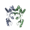
 Sample preparation
 Processing