 Movie
Movie Controller

[English] 日本語
 Yorodumi
- PDB-1ei1: DIMERIZATION OF E. COLI DNA GYRASE B PROVIDES A STRUCTURAL MECHAN... -
+ Open data

- Basic information
| Entry | Database: PDB / ID: 1ei1 | ||||||
|---|---|---|---|---|---|---|---|
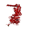
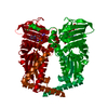
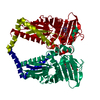
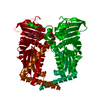
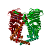
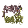
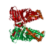
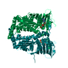
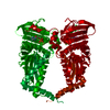
| Title | DIMERIZATION OF E. COLI DNA GYRASE B PROVIDES A STRUCTURAL MECHANISM FOR ACTIVATING THE ATPASE CATALYTIC CENTER | ||||||
 Components Components | DNA GYRASE B | ||||||
 Keywords Keywords | ISOMERASE / ATPase domain / dimer | ||||||
| Function / homology |  Function and homology information Function and homology informationAction of antimicrobials / DNA topoisomerase type II (double strand cut, ATP-hydrolyzing) complex / DNA negative supercoiling activity / DNA topoisomerase type II (double strand cut, ATP-hydrolyzing) activity / DNA topoisomerase (ATP-hydrolysing) / Antimicrobial resistance / DNA topological change / ATP-dependent activity, acting on DNA / DNA-templated DNA replication / chromosome ...Action of antimicrobials / DNA topoisomerase type II (double strand cut, ATP-hydrolyzing) complex / DNA negative supercoiling activity / DNA topoisomerase type II (double strand cut, ATP-hydrolyzing) activity / DNA topoisomerase (ATP-hydrolysing) / Antimicrobial resistance / DNA topological change / ATP-dependent activity, acting on DNA / DNA-templated DNA replication / chromosome / response to xenobiotic stimulus / response to antibiotic / DNA-templated transcription / DNA binding / ATP binding / metal ion binding / cytosol / cytoplasm Similarity search - Function | ||||||
| Biological species |  | ||||||
| Method |  X-RAY DIFFRACTION / SYNCHROTRON / Resolution: 2.3 Å X-RAY DIFFRACTION / SYNCHROTRON / Resolution: 2.3 Å | ||||||
 Authors Authors | Brino, L. / Urzhumtsev, A. / Oudet, P. / Moras, D. | ||||||
 Citation Citation | Journal: J.Biol.Chem. / Year: 2000 Title: Dimerization of Escherichia coli DNA-gyrase B provides a structural mechanism for activating the ATPase catalytic center. Authors: Brino, L. / Urzhumtsev, A. / Mousli, M. / Bronner, C. / Mitschler, A. / Oudet, P. / Moras, D. #1: Journal: Biochimie / Year: 1999Title: Isoleucine 10 is Essential for DNA Gyrase B Function in Escherichia coli. Authors: Brino, L. / Bronner, C. / Oudet, P. / Mousli, M. #2: Journal: Plasmid / Year: 1997Title: Expression in Escherichia coli of Y5-mutant and N-terminal Domain-deleted DNA Gyrase B Proteins Affects Strongly Plasmid Maintenance. Authors: Brino, L. / Mousli, M. / Oudet, P. / Weiss, E. | ||||||
| History |
|
- Structure visualization
| Structure viewer | Molecule: MolmilJmol/JSmol |
|---|
- Downloads & links
-Download
| PDBx/mmCIF format | 1ei1.cif.gz | 177.8 KB | Display | PDBx/mmCIF format |
|---|---|---|---|---|
| PDB format | pdb1ei1.ent.gz | 138 KB | Display | PDB format |
| PDBx/mmJSON format | 1ei1.json.gz | Tree view | PDBx/mmJSON format | |
| Others |  Other downloads Other downloads |
-Validation report
| Arichive directory | https://data.pdbj.org/pub/pdb/validation_reports/ei/1ei1ftp://data.pdbj.org/pub/pdb/validation_reports/ei/1ei1 | HTTPS FTP |
|---|
-Related structure data
| Similar structure data |
|---|

-Links
 PDBj
PDBj


- Assembly
| Deposited unit | 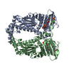
| ||||||||
|---|---|---|---|---|---|---|---|---|---|
| 1 |
| ||||||||
| Unit cell |
| ||||||||
| Details | The biological assembly is the dimer composed of chain A and B in the asymmetric unit. |
-Components
| #1: Protein | Mass: 43083.457 Da / Num. of mol.: 2 / Fragment: N-TERMINAL 43 KDA FRAGMENT / Mutation: Y5S, Y405S Source method: isolated from a genetically manipulated source Source: (gene. exp.) Description: PN43-Y5S (TAC PROMOTER, BETA LACTAMASE GENE, LAC IQ GENE, PBR322 BACKGROUND) Plasmid: PTTQ18 VECTOR(PAG111; PN43-Y5S) / Production host: References: UniProt: P06982, UniProt: P0AES6*PLUS, EC: 5.99.1.3 #2: Chemical |   Mass: 96.063 Da / Num. of mol.: 3 / Source method: obtained synthetically / Formula: SO4 Mass: 96.063 Da / Num. of mol.: 3 / Source method: obtained synthetically / Formula: SO4#3: Chemical |   Mass: 506.196 Da / Num. of mol.: 2 / Source method: obtained synthetically / Formula: C10H17N6O12P3 / Comment: AMP-PNP, energy-carrying molecule analogue*YM Mass: 506.196 Da / Num. of mol.: 2 / Source method: obtained synthetically / Formula: C10H17N6O12P3 / Comment: AMP-PNP, energy-carrying molecule analogue*YM#4: Chemical |   Mass: 92.094 Da / Num. of mol.: 2 / Source method: obtained synthetically / Formula: C3H8O3 Mass: 92.094 Da / Num. of mol.: 2 / Source method: obtained synthetically / Formula: C3H8O3#5: Water | ChemComp-HOH / |  Mass: 18.015 Da / Num. of mol.: 505 / Source method: isolated from a natural source / Formula: H2O Mass: 18.015 Da / Num. of mol.: 505 / Source method: isolated from a natural source / Formula: H2O |
|---|
-Experimental details
-Experiment
| Experiment | Method: X-RAY DIFFRACTION / Number of used crystals: 1 |
|---|
- Sample preparation
| Crystal | Density Matthews: 2.66 Å3/Da / Density % sol: 53.82 % | ||||||||||||||||||||||||||||||||||||
|---|---|---|---|---|---|---|---|---|---|---|---|---|---|---|---|---|---|---|---|---|---|---|---|---|---|---|---|---|---|---|---|---|---|---|---|---|---|
| Crystal grow | Temperature: 279 K / Method: vapor diffusion, hanging drop / pH: 7.5 Details: PEG400, ammonium sulfate, ADPNP, HEPES, pH 7.5, VAPOR DIFFUSION, HANGING DROP, temperature 279K | ||||||||||||||||||||||||||||||||||||
| Crystal grow | *PLUS Temperature: 4 ℃ | ||||||||||||||||||||||||||||||||||||
| Components of the solutions | *PLUS
|
-Data collection
| Diffraction | Mean temperature: 120 K |
|---|---|
| Diffraction source | Source: SYNCHROTRON / Site: LURE  / Beamline: DW32 / Wavelength: 0.91 / Beamline: DW32 / Wavelength: 0.91 |
| Detector | Type: MARRESEARCH / Detector: IMAGE PLATE |
| Radiation | Protocol: SINGLE WAVELENGTH / Monochromatic (M) / Laue (L): M / Scattering type: x-ray |
| Radiation wavelength | Wavelength: 0.91 Å / Relative weight: 1 |
| Reflection | Resolution: 2.3→15 Å / Num. obs: 33856 / % possible obs: 80 % / Observed criterion σ(F): 3 / Redundancy: 3.4 % / Rmerge(I) obs: 0.044 / Net I/σ(I): 26.4 |
| Reflection shell | Resolution: 2.3→2.5 Å / Num. unique all: 33856 / % possible all: 56 |
| Reflection | *PLUS Num. measured all: 114762 |
| Reflection shell | *PLUS % possible obs: 56 % |
- Processing
| Software |
| ||||||||||||||||||||
|---|---|---|---|---|---|---|---|---|---|---|---|---|---|---|---|---|---|---|---|---|---|
| Refinement | Resolution: 2.3→6 Å / σ(F): 2 / Stereochemistry target values: XPLOR standard
| ||||||||||||||||||||
| Refinement step | Cycle: LAST / Resolution: 2.3→6 Å
| ||||||||||||||||||||
| Refine LS restraints |
| ||||||||||||||||||||
| Software | *PLUS Name: X-PLOR / Version: 3.1 / Classification: refinement | ||||||||||||||||||||
| Refinement | *PLUS Highest resolution: 2.3 Å / Lowest resolution: 6 Å / σ(F): 2 / Rfactor obs: 0.171 | ||||||||||||||||||||
| Solvent computation | *PLUS | ||||||||||||||||||||
| Displacement parameters | *PLUS | ||||||||||||||||||||
| Refine LS restraints | *PLUS
|
