 Movie
Movie Controller

[English] 日本語
 Yorodumi
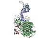
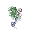
- PDB-2zc3: Penicillin-binding protein 2X (PBP 2X) acyl-enzyme complex (biape... -
+ Open data

- Basic information
| Entry | Database: PDB / ID: 2zc3 | ||||||
|---|---|---|---|---|---|---|---|
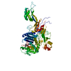
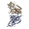
| Title | Penicillin-binding protein 2X (PBP 2X) acyl-enzyme complex (biapenem) from Streptococcus pneumoniae | ||||||
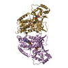
 Components Components | (Penicillin-binding protein ...) x 3 | ||||||
 Keywords Keywords | BIOSYNTHETIC PROTEIN / PEPTIDOGLYCAN SYNTHESIS / CELL WALL / PENICILLIN-BINDING / ANTIBIOTICS / BIAPENEM / Antibiotic resistance / Cell cycle / Cell division / Cell shape / Cell wall biogenesis/degradation / Membrane / Secreted / Transmembrane | ||||||
| Function / homology |  Function and homology information Function and homology informationpenicillin binding / peptidoglycan biosynthetic process / cell wall organization / regulation of cell shape / cell division / response to antibiotic / plasma membrane Similarity search - Function | ||||||
| Biological species |   Streptococcus pneumoniae (bacteria) Streptococcus pneumoniae (bacteria) | ||||||
| Method |  X-RAY DIFFRACTION / SYNCHROTRON / MOLECULAR REPLACEMENT / Resolution: 2.5 Å X-RAY DIFFRACTION / SYNCHROTRON / MOLECULAR REPLACEMENT / Resolution: 2.5 Å | ||||||
 Authors Authors | Yamada, M. / Watanabe, T. / Takeuchi, Y. | ||||||
 Citation Citation | Journal: Antimicrob.Agents Chemother. / Year: 2008 Title: Crystal Structures of Biapenem and Tebipenem Complexed with Penicillin-Binding Proteins 2X and 1A from Streptococcus pneumoniae Authors: Yamada, M. / Watanabe, T. / Baba, N. / Takeuchi, Y. / Ohsawa, F. / Gomi, S. | ||||||
| History |
|
- Structure visualization
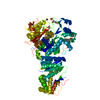
| Structure viewer | Molecule: MolmilJmol/JSmol |
|---|
- Downloads & links
-Download
| PDBx/mmCIF format | 2zc3.cif.gz | 252.8 KB | Display | PDBx/mmCIF format |
|---|---|---|---|---|
| PDB format | pdb2zc3.ent.gz | 202.8 KB | Display | PDB format |
| PDBx/mmJSON format | 2zc3.json.gz | Tree view | PDBx/mmJSON format | |
| Others |  Other downloads Other downloads |
-Validation report
| Arichive directory | https://data.pdbj.org/pub/pdb/validation_reports/zc/2zc3ftp://data.pdbj.org/pub/pdb/validation_reports/zc/2zc3 | HTTPS FTP |
|---|
-Related structure data
| Related structure data |  2zc4C 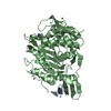 2zc5C 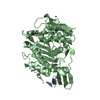 2zc6C  2z2lS C: citing same article ( S: Starting model for refinement |
|---|---|
| Similar structure data |


-Links
 PDBj
PDBj- Assembly
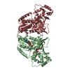
| Deposited unit | 
| ||||||||
|---|---|---|---|---|---|---|---|---|---|
| 1 | 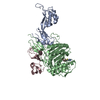
| ||||||||
| 2 | 
| ||||||||
| 3 | 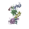
| ||||||||
| Unit cell |
|
-Components
-Penicillin-binding protein ... , 3 types, 6 molecules ADBECF
| #1: Protein | Mass: 18473.584 Da / Num. of mol.: 2 / Fragment: UNP residues 71-238 Source method: isolated from a genetically manipulated source Details: N-terminal domain / Source: (gene. exp.) Streptococcus pneumoniae (bacteria) / Gene: pbpX / Plasmid: pET15b / Production host: #2: Protein | Mass: 42000.016 Da / Num. of mol.: 2 / Fragment: UNP residues 241-625 Source method: isolated from a genetically manipulated source Details: Transpeptidase domain / Source: (gene. exp.) Streptococcus pneumoniae (bacteria) / Gene: pbpX / Plasmid: pET15b / Production host: #3: Protein | Mass: 13707.433 Da / Num. of mol.: 2 / Fragment: UNP residues 626-750 Source method: isolated from a genetically manipulated source Details: C-terminal domain / Source: (gene. exp.) Streptococcus pneumoniae (bacteria) / Gene: pbpX / Plasmid: pET15b / Production host: |
|---|
-Non-polymers , 3 types, 85 molecules 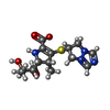


| #4: Chemical |  Mass: 352.409 Da / Num. of mol.: 2 / Source method: obtained synthetically / Formula: C15H20N4O4S Mass: 352.409 Da / Num. of mol.: 2 / Source method: obtained synthetically / Formula: C15H20N4O4S#5: Chemical | ChemComp-SO4 / |  Mass: 96.063 Da / Num. of mol.: 1 / Source method: obtained synthetically / Formula: SO4 Mass: 96.063 Da / Num. of mol.: 1 / Source method: obtained synthetically / Formula: SO4#6: Water | ChemComp-HOH / | Mass: 18.015 Da / Num. of mol.: 82 / Source method: isolated from a natural source / Formula: H2O |
|---|
-Details
| Has protein modification | Y |
|---|
-Experimental details
-Experiment
| Experiment | Method: X-RAY DIFFRACTION / Number of used crystals: 1 |
|---|
- Sample preparation
| Crystal | Density Matthews: 2.74 Å3/Da / Density % sol: 55.16 % |
|---|---|
| Crystal grow | Temperature: 293 K / Method: vapor diffusion, hanging drop Details: 10-25% PEG4000, 0.2M ammonium sulfate, 0.1M sodium acetate, VAPOR DIFFUSION, HANGING DROP, temperature 293K |
-Data collection
| Diffraction | Mean temperature: 100 K |
|---|---|
| Diffraction source | Source: SYNCHROTRON / Site: SPring-8  / Beamline: BL32B2 / Wavelength: 1 Å / Beamline: BL32B2 / Wavelength: 1 Å |
| Detector | Type: RIGAKU RAXIS V / Detector: IMAGE PLATE / Date: Mar 16, 2006 Details: A fixed exit SI double crystal monochromator followed bystandard double crystal monochromator and RH-coated downward-deflection mirror with a typical glancing angle of 3.7MRAD |
| Radiation | Protocol: SINGLE WAVELENGTH / Monochromatic (M) / Laue (L): M / Scattering type: x-ray |
| Radiation wavelength | Wavelength: 1 Å / Relative weight: 1 |
| Reflection | Resolution: 2.5→29.51 Å / Num. obs: 53714 / % possible obs: 92.7 % / Redundancy: 5.3 % / Biso Wilson estimate: 52.76 Å2 / Rmerge(I) obs: 0.059 / Net I/σ(I): 18.9 |
| Reflection shell | Resolution: 2.5→2.58 Å / Redundancy: 4.8 % / Rmerge(I) obs: 0.228 / Mean I/σ(I) obs: 5.5 / % possible all: 90.1 |
- Processing
| Software |
| ||||||||||||||||||||||||||||||||||||||||||||||||||||||||||||||||||||||||||||||||||||||||||||||||||||||||||||||||||||||||||||||||||||||||||||||||||||||||||||||||||||||||||
|---|---|---|---|---|---|---|---|---|---|---|---|---|---|---|---|---|---|---|---|---|---|---|---|---|---|---|---|---|---|---|---|---|---|---|---|---|---|---|---|---|---|---|---|---|---|---|---|---|---|---|---|---|---|---|---|---|---|---|---|---|---|---|---|---|---|---|---|---|---|---|---|---|---|---|---|---|---|---|---|---|---|---|---|---|---|---|---|---|---|---|---|---|---|---|---|---|---|---|---|---|---|---|---|---|---|---|---|---|---|---|---|---|---|---|---|---|---|---|---|---|---|---|---|---|---|---|---|---|---|---|---|---|---|---|---|---|---|---|---|---|---|---|---|---|---|---|---|---|---|---|---|---|---|---|---|---|---|---|---|---|---|---|---|---|---|---|---|---|---|---|---|
| Refinement | Method to determine structure: MOLECULAR REPLACEMENT Starting model: PDB ENTRY 2Z2L Resolution: 2.5→29.51 Å / Cor.coef. Fo:Fc: 0.928 / Cor.coef. Fo:Fc free: 0.893 / SU B: 11.417 / SU ML: 0.248 / Cross valid method: THROUGHOUT / ESU R: 0.594 / ESU R Free: 0.326 / Stereochemistry target values: MAXIMUM LIKELIHOOD / Details: Hydrogens have been added in the riding positions
| ||||||||||||||||||||||||||||||||||||||||||||||||||||||||||||||||||||||||||||||||||||||||||||||||||||||||||||||||||||||||||||||||||||||||||||||||||||||||||||||||||||||||||
| Solvent computation | Ion probe radii: 0.8 Å / Shrinkage radii: 0.8 Å / VDW probe radii: 1.2 Å / Solvent model: MASK | ||||||||||||||||||||||||||||||||||||||||||||||||||||||||||||||||||||||||||||||||||||||||||||||||||||||||||||||||||||||||||||||||||||||||||||||||||||||||||||||||||||||||||
| Displacement parameters | Biso mean: 47.99 Å2
| ||||||||||||||||||||||||||||||||||||||||||||||||||||||||||||||||||||||||||||||||||||||||||||||||||||||||||||||||||||||||||||||||||||||||||||||||||||||||||||||||||||||||||
| Refinement step | Cycle: LAST / Resolution: 2.5→29.51 Å
| ||||||||||||||||||||||||||||||||||||||||||||||||||||||||||||||||||||||||||||||||||||||||||||||||||||||||||||||||||||||||||||||||||||||||||||||||||||||||||||||||||||||||||
| Refine LS restraints |
| ||||||||||||||||||||||||||||||||||||||||||||||||||||||||||||||||||||||||||||||||||||||||||||||||||||||||||||||||||||||||||||||||||||||||||||||||||||||||||||||||||||||||||
| LS refinement shell | Resolution: 2.5→2.564 Å / Total num. of bins used: 20
|
