 Movie
Movie Controller

[English] 日本語
 Yorodumi
- PDB-2z2m: Cefditoren-Acylated Penicillin-Binding Protein 2X (PBP2X) from St... -
+ Open data

- Basic information
| Entry | Database: PDB / ID: 2z2m | ||||||
|---|---|---|---|---|---|---|---|
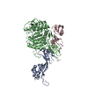
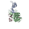
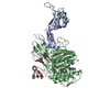
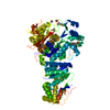
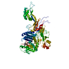
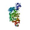
| Title | Cefditoren-Acylated Penicillin-Binding Protein 2X (PBP2X) from Streptococcus pneumoniae | ||||||
 Components Components | (Penicillin-binding protein ...) x 3 | ||||||
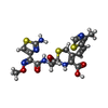
 Keywords Keywords | BIOSYNTHETIC PROTEIN / PEPTIDOGLYCAN SYNTHESIS / CELL WALL / PENICILLIN-BINDING / ANTIBIOTICS / CEFDITOREN | ||||||
| Function / homology |  Function and homology information Function and homology informationpenicillin binding / peptidoglycan biosynthetic process / cell wall organization / regulation of cell shape / cell division / response to antibiotic / plasma membrane Similarity search - Function | ||||||
| Biological species |   Streptococcus pneumoniae (bacteria) Streptococcus pneumoniae (bacteria) | ||||||
| Method |  X-RAY DIFFRACTION / SYNCHROTRON / MOLECULAR REPLACEMENT / Resolution: 2.6 Å X-RAY DIFFRACTION / SYNCHROTRON / MOLECULAR REPLACEMENT / Resolution: 2.6 Å | ||||||
 Authors Authors | Yamada, M. / Watanabe, T. / Takeuchi, Y. | ||||||
 Citation Citation | Journal: Antimicrob.Agents Chemother. / Year: 2007 Title: Crystal Structure of Cefditoren Complexed with Streptococcus pneumoniae Penicillin-Binding Protein 2X: Structural Basis for its High Antimicrobial Activity Authors: Yamada, M. / Watanabe, T. / Miyara, T. / Baba, N. / Saito, J. / Takeuchi, Y. / Ohsawa, F. | ||||||
| History |
|
- Structure visualization
| Structure viewer | Molecule: MolmilJmol/JSmol |
|---|
- Downloads & links
-Download
| PDBx/mmCIF format | 2z2m.cif.gz | 243 KB | Display | PDBx/mmCIF format |
|---|---|---|---|---|
| PDB format | pdb2z2m.ent.gz | 194.9 KB | Display | PDB format |
| PDBx/mmJSON format | 2z2m.json.gz | Tree view | PDBx/mmJSON format | |
| Others |  Other downloads Other downloads |
-Validation report
| Arichive directory | https://data.pdbj.org/pub/pdb/validation_reports/z2/2z2mftp://data.pdbj.org/pub/pdb/validation_reports/z2/2z2m | HTTPS FTP |
|---|
-Related structure data
| Related structure data |  2z2lSC S: Starting model for refinement C: citing same article ( |
|---|---|
| Similar structure data |


-Links
 PDBj
PDBj- Assembly
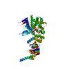
| Deposited unit | 
| ||||||||
|---|---|---|---|---|---|---|---|---|---|
| 1 | 
| ||||||||
| 2 | 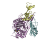
| ||||||||
| Unit cell |
|
-Components
-Penicillin-binding protein ... , 3 types, 6 molecules ADBECF
| #1: Protein | Mass: 18473.584 Da / Num. of mol.: 2 / Fragment: UNP residues 71-238 Source method: isolated from a genetically manipulated source Details: N-TERMINAL DOMAIN / Source: (gene. exp.) Streptococcus pneumoniae (bacteria) / Gene: pbpX / Plasmid: pET15b / Production host: #2: Protein | Mass: 42000.016 Da / Num. of mol.: 2 / Fragment: UNP residues 241-625 Source method: isolated from a genetically manipulated source Details: TRANSPEPTIDASE DOMAIN / Source: (gene. exp.) Streptococcus pneumoniae (bacteria) / Gene: pbpX / Plasmid: pET15b / Production host: #3: Protein | Mass: 13707.433 Da / Num. of mol.: 2 / Fragment: UNP residues 626-750 Source method: isolated from a genetically manipulated source Details: C-TERMINAL DOMAIN / Source: (gene. exp.) Streptococcus pneumoniae (bacteria) / Gene: pbpX / Plasmid: pET15b / Production host: |
|---|
-Non-polymers , 3 types, 96 molecules 

| #4: Chemical | ChemComp-SO4 /  Mass: 96.063 Da / Num. of mol.: 1 / Source method: obtained synthetically / Formula: SO4 Mass: 96.063 Da / Num. of mol.: 1 / Source method: obtained synthetically / Formula: SO4 | ||
|---|---|---|---|
| #5: Chemical |  Mass: 508.594 Da / Num. of mol.: 2 / Source method: obtained synthetically / Formula: C19H20N6O5S3 Mass: 508.594 Da / Num. of mol.: 2 / Source method: obtained synthetically / Formula: C19H20N6O5S3#6: Water | ChemComp-HOH / | Mass: 18.015 Da / Num. of mol.: 93 / Source method: isolated from a natural source / Formula: H2O |
-Details
| Has protein modification | Y |
|---|
-Experimental details
-Experiment
| Experiment | Method: X-RAY DIFFRACTION / Number of used crystals: 1 |
|---|
- Sample preparation
| Crystal | Density Matthews: 2.77 Å3/Da / Density % sol: 55.59 % |
|---|---|
| Crystal grow | Method: vapor diffusion, hanging drop Details: 10-25% PEG4000, 0.2M ammonium sulfate, 0.1M sodium acetate, VAPOR DIFFUSION, HANGING DROP |
-Data collection
| Diffraction | Mean temperature: 100 K |
|---|---|
| Diffraction source | Source: SYNCHROTRON / Site: SPring-8  / Beamline: BL32B2 / Wavelength: 1 Å / Beamline: BL32B2 / Wavelength: 1 Å |
| Detector | Type: RIGAKU JUPITER 210 / Detector: CCD / Date: Nov 11, 2005 |
| Radiation | Protocol: SINGLE WAVELENGTH / Monochromatic (M) / Laue (L): M / Scattering type: x-ray |
| Radiation wavelength | Wavelength: 1 Å / Relative weight: 1 |
| Reflection | Resolution: 2.6→30 Å / Num. obs: 46229 / % possible obs: 89.9 % / Redundancy: 4.3 % / Biso Wilson estimate: 60.179 Å2 / Rmerge(I) obs: 0.054 / Net I/σ(I): 16.7 |
| Reflection shell | Resolution: 2.6→2.69 Å / Redundancy: 4 % / Rmerge(I) obs: 0.215 / Mean I/σ(I) obs: 5.5 / % possible all: 90.5 |
- Processing
| Software |
| ||||||||||||||||||||||||||||||||||||||||||||||||||||||||||||||||||||||||||||||||||||||||||||||||||||||||||||||||||||||||||||||||||||||||||||||||||||||||||||||||||||||||||
|---|---|---|---|---|---|---|---|---|---|---|---|---|---|---|---|---|---|---|---|---|---|---|---|---|---|---|---|---|---|---|---|---|---|---|---|---|---|---|---|---|---|---|---|---|---|---|---|---|---|---|---|---|---|---|---|---|---|---|---|---|---|---|---|---|---|---|---|---|---|---|---|---|---|---|---|---|---|---|---|---|---|---|---|---|---|---|---|---|---|---|---|---|---|---|---|---|---|---|---|---|---|---|---|---|---|---|---|---|---|---|---|---|---|---|---|---|---|---|---|---|---|---|---|---|---|---|---|---|---|---|---|---|---|---|---|---|---|---|---|---|---|---|---|---|---|---|---|---|---|---|---|---|---|---|---|---|---|---|---|---|---|---|---|---|---|---|---|---|---|---|---|
| Refinement | Method to determine structure: MOLECULAR REPLACEMENT Starting model: 2Z2L Resolution: 2.6→29.42 Å / Cor.coef. Fo:Fc: 0.93 / Cor.coef. Fo:Fc free: 0.898 / SU B: 10.142 / SU ML: 0.22 / Cross valid method: THROUGHOUT / ESU R: 0.805 / ESU R Free: 0.343 / Stereochemistry target values: MAXIMUM LIKELIHOOD / Details: HYDROGENS HAVE BEEN ADDED IN THE RIDING POSITIONS
| ||||||||||||||||||||||||||||||||||||||||||||||||||||||||||||||||||||||||||||||||||||||||||||||||||||||||||||||||||||||||||||||||||||||||||||||||||||||||||||||||||||||||||
| Solvent computation | Ion probe radii: 0.8 Å / Shrinkage radii: 0.8 Å / VDW probe radii: 1.2 Å / Solvent model: MASK | ||||||||||||||||||||||||||||||||||||||||||||||||||||||||||||||||||||||||||||||||||||||||||||||||||||||||||||||||||||||||||||||||||||||||||||||||||||||||||||||||||||||||||
| Displacement parameters | Biso mean: 46.402 Å2
| ||||||||||||||||||||||||||||||||||||||||||||||||||||||||||||||||||||||||||||||||||||||||||||||||||||||||||||||||||||||||||||||||||||||||||||||||||||||||||||||||||||||||||
| Refinement step | Cycle: LAST / Resolution: 2.6→29.42 Å
| ||||||||||||||||||||||||||||||||||||||||||||||||||||||||||||||||||||||||||||||||||||||||||||||||||||||||||||||||||||||||||||||||||||||||||||||||||||||||||||||||||||||||||
| Refine LS restraints |
| ||||||||||||||||||||||||||||||||||||||||||||||||||||||||||||||||||||||||||||||||||||||||||||||||||||||||||||||||||||||||||||||||||||||||||||||||||||||||||||||||||||||||||
| LS refinement shell | Resolution: 2.6→2.667 Å / Total num. of bins used: 20
|
