 Movie
Movie Controller

[English] 日本語
 Yorodumi
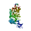
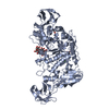
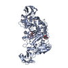
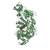
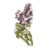
- PDB-3wdi: Crystal structure of Pullulanase complexed with maltotriose from ... -
+ Open data

- Basic information
| Entry | Database: PDB / ID: 3wdi | |||||||||
|---|---|---|---|---|---|---|---|---|---|---|
| Title | Crystal structure of Pullulanase complexed with maltotriose from Anoxybacillus sp. LM18-11 | |||||||||
 Components Components | Type I pullulanase | |||||||||
 Keywords Keywords | HYDROLASE / glycoside hydrolase / pullulanase | |||||||||
| Function / homology |  Function and homology information Function and homology informationpullulanase / pullulanase activity / carbohydrate metabolic process / metal ion binding Similarity search - Function | |||||||||
| Biological species |  Anoxybacillus sp. LM18-11 (bacteria) Anoxybacillus sp. LM18-11 (bacteria) | |||||||||
| Method |  X-RAY DIFFRACTION / SYNCHROTRON / MOLECULAR REPLACEMENT / Resolution: 2.2 Å X-RAY DIFFRACTION / SYNCHROTRON / MOLECULAR REPLACEMENT / Resolution: 2.2 Å | |||||||||
 Authors Authors | Xu, J. / Ren, F. / Huang, C.H. / Zheng, Y. / Zhen, J. / Ko, T.P. / Chen, C.C. / Chan, H.C. / Guo, R.T. / Ma, Y. / Song, H. | |||||||||
 Citation Citation | Journal: To be Published Title: Cloning, Expression, Functional and Structural Studies of Pullulanase from Anoxybacillus sp. LM18-11 Authors: Xu, J. / Ren, F. / Huang, C.H. / Zheng, Y. / Zhen, J. / Chen, C.C. / Chan, H.C. / Guo, R.T. / Ma, Y. / Song, H. | |||||||||
| History |
|
- Structure visualization
| Structure viewer | Molecule: MolmilJmol/JSmol |
|---|
- Downloads & links
-Download
| PDBx/mmCIF format | 3wdi.cif.gz | 171.2 KB | Display | PDBx/mmCIF format |
|---|---|---|---|---|
| PDB format | pdb3wdi.ent.gz | 133 KB | Display | PDB format |
| PDBx/mmJSON format | 3wdi.json.gz | Tree view | PDBx/mmJSON format | |
| Others |  Other downloads Other downloads |
-Validation report
| Arichive directory | https://data.pdbj.org/pub/pdb/validation_reports/wd/3wdiftp://data.pdbj.org/pub/pdb/validation_reports/wd/3wdi | HTTPS FTP |
|---|
-Related structure data
| Related structure data |  3wdhSC 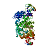 3wdjC S: Starting model for refinement C: citing same article ( |
|---|---|
| Similar structure data |


-Links
 PDBj
PDBj

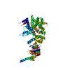
- Assembly
| Deposited unit | 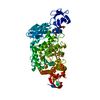
| ||||||||||||
|---|---|---|---|---|---|---|---|---|---|---|---|---|---|
| 1 |
| ||||||||||||
| Unit cell |
| ||||||||||||
| Components on special symmetry positions |
|
-Components
| #1: Protein | Mass: 82172.422 Da / Num. of mol.: 1 / Fragment: UNP residues 3-707 Source method: isolated from a genetically manipulated source Source: (gene. exp.) Anoxybacillus sp. LM18-11 (bacteria) / Gene: pulA / Plasmid: pET32a / Production host: | ||||
|---|---|---|---|---|---|
| #2: Polysaccharide | alpha-D-glucopyranose-(1-4)-alpha-D-glucopyranose-(1-4)-alpha-D-glucopyranose / alpha-maltotriose   Source method: isolated from a genetically manipulated source Details: oligosaccharide / References: alpha-maltotriose #3: Chemical | ChemComp-CA / |   Mass: 40.078 Da / Num. of mol.: 1 / Source method: obtained synthetically / Formula: Ca Mass: 40.078 Da / Num. of mol.: 1 / Source method: obtained synthetically / Formula: Ca#4: Water | ChemComp-HOH / |  Mass: 18.015 Da / Num. of mol.: 555 / Source method: isolated from a natural source / Formula: H2O Mass: 18.015 Da / Num. of mol.: 555 / Source method: isolated from a natural source / Formula: H2O |
-Experimental details
-Experiment
| Experiment | Method: X-RAY DIFFRACTION / Number of used crystals: 1 |
|---|
- Sample preparation
| Crystal | Density Matthews: 2.54 Å3/Da / Density % sol: 51.52 % |
|---|---|
| Crystal grow | Temperature: 295 K / pH: 7.5 Details: 0.1M HEPES, pH 7.5, 7%(w/v) Polyethylene Glycol 8000, 10%(v/v) Ethylene Glycol , VAPOR DIFFUSION, SITTING DROP, temperature 295K |
-Data collection
| Diffraction | Mean temperature: 100 K |
|---|---|
| Diffraction source | Source: SYNCHROTRON / Site: NSRRC  / Beamline: BL13B1 / Wavelength: 1 / Beamline: BL13B1 / Wavelength: 1 |
| Detector | Type: ADSC QUANTUM 315 / Detector: CCD / Date: Jun 20, 2011 |
| Radiation | Protocol: SINGLE WAVELENGTH / Monochromatic (M) / Laue (L): M / Scattering type: x-ray |
| Radiation wavelength | Wavelength: 1 Å / Relative weight: 1 |
| Reflection | Resolution: 2.2→25 Å / Num. obs: 43239 / % possible obs: 99.7 % / Redundancy: 7.2 % / Rmerge(I) obs: 0.082 / Net I/σ(I): 26.6 |
| Reflection shell | Resolution: 2.2→2.28 Å / Redundancy: 7.2 % / Rmerge(I) obs: 0.475 / Mean I/σ(I) obs: 4.5 / % possible all: 99.3 |
- Processing
| Software |
| ||||||||||||||||||||
|---|---|---|---|---|---|---|---|---|---|---|---|---|---|---|---|---|---|---|---|---|---|
| Refinement | Method to determine structure: MOLECULAR REPLACEMENT Starting model: 3WDH Resolution: 2.2→25 Å / σ(F): 2 / Stereochemistry target values: Engh & Huber
| ||||||||||||||||||||
| Refinement step | Cycle: LAST / Resolution: 2.2→25 Å
| ||||||||||||||||||||
| LS refinement shell | Resolution: 2.2→2.28 Å /
|
