 Movie
Movie Controller

[English] 日本語
 Yorodumi
- PDB-2zbd: Crystal Structure of the SR Calcium Pump with Bound Aluminium Flu... -
+ Open data

- Basic information
| Entry | Database: PDB / ID: 2zbd | |||||||||
|---|---|---|---|---|---|---|---|---|---|---|
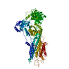
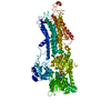
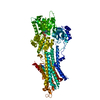
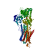
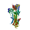
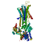
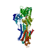
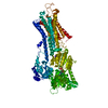
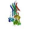
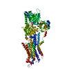
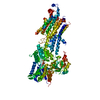
| Title | Crystal Structure of the SR Calcium Pump with Bound Aluminium Fluoride, ADP and Calcium | |||||||||
 Components Components | Sarcoplasmic/endoplasmic reticulum calcium ATPase 1 | |||||||||
 Keywords Keywords | HYDROLASE / MEMBRANE PROTEIN / P-TYPE ATPASE / HAD FOLD / ALTERNATIVE SPLICING / ATP-BINDING / CALCIUM / CALCIUM TRANSPORT / ENDOPLASMIC RETICULUM / ION TRANSPORT / MAGNESIUM / METAL-BINDING / NUCLEOTIDE-BINDING / PHOSPHORYLATION / SARCOPLASMIC RETICULUM / TRANSMEMBRANE / TRANSPORT | |||||||||
| Function / homology |  Function and homology information Function and homology informationpositive regulation of calcium ion import into sarcoplasmic reticulum / positive regulation of ATPase-coupled calcium transmembrane transporter activity / positive regulation of fast-twitch skeletal muscle fiber contraction / H zone / calcium ion import into sarcoplasmic reticulum / negative regulation of striated muscle contraction / regulation of striated muscle contraction / P-type Ca2+ transporter / P-type calcium transporter activity / positive regulation of cardiac muscle cell contraction ...positive regulation of calcium ion import into sarcoplasmic reticulum / positive regulation of ATPase-coupled calcium transmembrane transporter activity / positive regulation of fast-twitch skeletal muscle fiber contraction / H zone / calcium ion import into sarcoplasmic reticulum / negative regulation of striated muscle contraction / regulation of striated muscle contraction / P-type Ca2+ transporter / P-type calcium transporter activity / positive regulation of cardiac muscle cell contraction / I band / endoplasmic reticulum-Golgi intermediate compartment / sarcoplasmic reticulum membrane / sarcoplasmic reticulum / calcium ion transport / calcium ion binding / endoplasmic reticulum membrane / perinuclear region of cytoplasm / endoplasmic reticulum / ATP hydrolysis activity / ATP binding / membrane Similarity search - Function | |||||||||
| Biological species |  | |||||||||
| Method |  X-RAY DIFFRACTION / SYNCHROTRON / MOLECULAR REPLACEMENT / Resolution: 2.4 Å X-RAY DIFFRACTION / SYNCHROTRON / MOLECULAR REPLACEMENT / Resolution: 2.4 Å | |||||||||
 Authors Authors | Toyoshima, C. / Nomura, H. / Tsuda, T. / Ogawa, H. / Norimatsu, Y. | |||||||||
 Citation Citation | Journal: Nature / Year: 2004 Title: Lumenal gating mechanism revealed in calcium pump crystal structures with phosphate analogues Authors: Toyoshima, C. / Nomura, H. / Tsuda, T. #1: Journal: Nature / Year: 2000Title: Crystal structure of the calcium pump of sarcoplasmic reticulum at 2.6 A resolution Authors: Toyoshima, C. / Nakasako, M. / Nomura, H. / Ogawa, H. #2: Journal: Nature / Year: 2002Title: Structural changes in the calcium pump accompanying the dissociation of calcium Authors: Toyoshima, C. / Nomura, H. #3: Journal: Nature / Year: 2004Title: Crystal structure of the calcium pump with a bound ATP analogue Authors: Toyoshima, C. / Mizutani, T. #4: Journal: Proc.Natl.Acad.Sci.Usa / Year: 2005Title: Structural role of countertransport revealed in Ca(2+) pump crystal structure in the absence of Ca(2+) Authors: Obara, K. / Miyashita, N. / Xu, C. / Toyoshima, I. / Sugita, Y. / Inesi, G. / Toyoshima, C. #5: Journal: Proc.Natl.Acad.Sci.Usa / Year: 2007Title: Interdomain communication in calcium pump as revealed in the crystal structures with transmembrane inhibitors Authors: Takahashi, M. / Kondou, Y. / Toyoshima, C. | |||||||||
| History |
| |||||||||
| Remark 999 | SEQUENCE The C-terminal residues in UNP entry P04191 are from 994 to 1001, DPEDERRK. In isoform ...SEQUENCE The C-terminal residues in UNP entry P04191 are from 994 to 1001, DPEDERRK. In isoform SERCA1A, there is only one c-terminal residue 994 GLY. |
- Structure visualization
| Structure viewer | Molecule: MolmilJmol/JSmol |
|---|
- Downloads & links
-Download
| PDBx/mmCIF format | 2zbd.cif.gz | 210.5 KB | Display | PDBx/mmCIF format |
|---|---|---|---|---|
| PDB format | pdb2zbd.ent.gz | 164.7 KB | Display | PDB format |
| PDBx/mmJSON format | 2zbd.json.gz | Tree view | PDBx/mmJSON format | |
| Others |  Other downloads Other downloads |
-Validation report
| Arichive directory | https://data.pdbj.org/pub/pdb/validation_reports/zb/2zbdftp://data.pdbj.org/pub/pdb/validation_reports/zb/2zbd | HTTPS FTP |
|---|
-Related structure data
| Related structure data |  1wpgC  1wpe C: citing same article ( S: Starting model for refinement |
|---|---|
| Similar structure data |


-Links
 PDBj
PDBj


- Assembly
| Deposited unit | 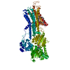
| ||||||||
|---|---|---|---|---|---|---|---|---|---|
| 1 |
| ||||||||
| Unit cell |
|
-Components
-Protein , 1 types, 1 molecules A
| #1: Protein | Mass: 109628.617 Da / Num. of mol.: 1 / Source method: isolated from a natural source / Source: (natural) |
|---|
-Non-polymers , 6 types, 106 molecules 


| #2: Chemical |  Mass: 40.078 Da / Num. of mol.: 2 / Source method: obtained synthetically / Formula: Ca Mass: 40.078 Da / Num. of mol.: 2 / Source method: obtained synthetically / Formula: Ca#3: Chemical |  Mass: 24.305 Da / Num. of mol.: 2 / Source method: obtained synthetically / Formula: Mg Mass: 24.305 Da / Num. of mol.: 2 / Source method: obtained synthetically / Formula: Mg#4: Chemical | ChemComp-ALF / |  Mass: 102.975 Da / Num. of mol.: 1 / Source method: obtained synthetically / Formula: AlF4 Mass: 102.975 Da / Num. of mol.: 1 / Source method: obtained synthetically / Formula: AlF4#5: Chemical | ChemComp-ADP / |  Mass: 427.201 Da / Num. of mol.: 1 / Source method: obtained synthetically / Formula: C10H15N5O10P2 / Comment: ADP, energy-carrying molecule*YM Mass: 427.201 Da / Num. of mol.: 1 / Source method: obtained synthetically / Formula: C10H15N5O10P2 / Comment: ADP, energy-carrying molecule*YM#6: Chemical |  Mass: 790.145 Da / Num. of mol.: 2 / Source method: obtained synthetically / Formula: C44H88NO8P / Comment: phospholipid*YM Mass: 790.145 Da / Num. of mol.: 2 / Source method: obtained synthetically / Formula: C44H88NO8P / Comment: phospholipid*YM#7: Water | ChemComp-HOH / | Mass: 18.015 Da / Num. of mol.: 98 / Source method: isolated from a natural source / Formula: H2O |
|---|
-Details
| Has protein modification | Y |
|---|
-Experimental details
-Experiment
| Experiment | Method: X-RAY DIFFRACTION / Number of used crystals: 1 |
|---|
- Sample preparation
| Crystal | Density Matthews: 4.01 Å3/Da / Density % sol: 69.29 % |
|---|---|
| Crystal grow | Temperature: 283 K / pH: 6.1 / Details: PEG 400, pH 6.10, MICRODIALYSIS, temperature 283K |
-Data collection
| Diffraction | Mean temperature: 100 K |
|---|---|
| Diffraction source | Source: SYNCHROTRON / Site: SPring-8  / Beamline: BL41XU / Wavelength: 0.8 / Beamline: BL41XU / Wavelength: 0.8 |
| Detector | Type: ADSC QUANTUM 315 / Detector: CCD / Date: Jul 13, 2005 |
| Radiation | Monochromator: ROTATED-INCLINED DOUBLE- CRYSTAL / Protocol: SINGLE WAVELENGTH / Monochromatic (M) / Laue (L): M / Scattering type: x-ray |
| Radiation wavelength | Wavelength: 0.8 Å / Relative weight: 1 |
| Reflection | Resolution: 2.4→20 Å / Num. obs: 61807 / % possible obs: 90.8 % / Observed criterion σ(I): -3 / Redundancy: 3.5 % / Rmerge(I) obs: 0.054 / Net I/σ(I): 27.7 |
| Reflection shell | Resolution: 2.4→2.46 Å / Redundancy: 2.9 % / Rmerge(I) obs: 0.35 / Mean I/σ(I) obs: 2.7 / % possible all: 58.5 |
- Processing
| Software |
| |||||||||||||||||||||||||||||||||||||||||||||||||||||||||||||||||||||||||||||||||||||||||||||||||||||||||||||||||||||||||||||||||||||||||||||||||||||||||||||||||||||||||||||||
|---|---|---|---|---|---|---|---|---|---|---|---|---|---|---|---|---|---|---|---|---|---|---|---|---|---|---|---|---|---|---|---|---|---|---|---|---|---|---|---|---|---|---|---|---|---|---|---|---|---|---|---|---|---|---|---|---|---|---|---|---|---|---|---|---|---|---|---|---|---|---|---|---|---|---|---|---|---|---|---|---|---|---|---|---|---|---|---|---|---|---|---|---|---|---|---|---|---|---|---|---|---|---|---|---|---|---|---|---|---|---|---|---|---|---|---|---|---|---|---|---|---|---|---|---|---|---|---|---|---|---|---|---|---|---|---|---|---|---|---|---|---|---|---|---|---|---|---|---|---|---|---|---|---|---|---|---|---|---|---|---|---|---|---|---|---|---|---|---|---|---|---|---|---|---|---|---|
| Refinement | Method to determine structure: MOLECULAR REPLACEMENT Starting model: PDB ENTRY 1WPE 1wpe Resolution: 2.4→12 Å / Cor.coef. Fo:Fc: 0.94 / Cor.coef. Fo:Fc free: 0.929 / SU B: 22.182 / SU ML: 0.246 / Cross valid method: THROUGHOUT / ESU R: 0.328 / ESU R Free: 0.244 / Stereochemistry target values: MAXIMUM LIKELIHOOD
| |||||||||||||||||||||||||||||||||||||||||||||||||||||||||||||||||||||||||||||||||||||||||||||||||||||||||||||||||||||||||||||||||||||||||||||||||||||||||||||||||||||||||||||||
| Solvent computation | Ion probe radii: 0.8 Å / Shrinkage radii: 0.8 Å / VDW probe radii: 1.4 Å / Solvent model: BABINET MODEL WITH MASK | |||||||||||||||||||||||||||||||||||||||||||||||||||||||||||||||||||||||||||||||||||||||||||||||||||||||||||||||||||||||||||||||||||||||||||||||||||||||||||||||||||||||||||||||
| Displacement parameters | Biso mean: 92.569 Å2
| |||||||||||||||||||||||||||||||||||||||||||||||||||||||||||||||||||||||||||||||||||||||||||||||||||||||||||||||||||||||||||||||||||||||||||||||||||||||||||||||||||||||||||||||
| Refinement step | Cycle: LAST / Resolution: 2.4→12 Å
| |||||||||||||||||||||||||||||||||||||||||||||||||||||||||||||||||||||||||||||||||||||||||||||||||||||||||||||||||||||||||||||||||||||||||||||||||||||||||||||||||||||||||||||||
| Refine LS restraints |
| |||||||||||||||||||||||||||||||||||||||||||||||||||||||||||||||||||||||||||||||||||||||||||||||||||||||||||||||||||||||||||||||||||||||||||||||||||||||||||||||||||||||||||||||
| LS refinement shell | Resolution: 2.4→2.46 Å / Total num. of bins used: 20
| |||||||||||||||||||||||||||||||||||||||||||||||||||||||||||||||||||||||||||||||||||||||||||||||||||||||||||||||||||||||||||||||||||||||||||||||||||||||||||||||||||||||||||||||
| Refinement TLS params. | Method: refined / Refine-ID: X-RAY DIFFRACTION
| |||||||||||||||||||||||||||||||||||||||||||||||||||||||||||||||||||||||||||||||||||||||||||||||||||||||||||||||||||||||||||||||||||||||||||||||||||||||||||||||||||||||||||||||
| Refinement TLS group |
|
