 Movie
Movie Controller


+ Open data

- Basic information
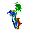
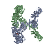
| Entry | Database: PDB / ID: 1wpg | ||||||
|---|---|---|---|---|---|---|---|
| Title | Crystal structure of the SR CA2+-ATPase with MGF4 | ||||||
 Components Components | Sarcoplasmic/endoplasmic reticulum calcium ATPase 1 | ||||||
 Keywords Keywords | HYDROLASE/HYDROLASE INHIBITOR / MEMBRANE PROTEIN / P-TYPE ATPASE / HAD FOLD / HYDROLASE / HYDROLASE-HYDROLASE INHIBITOR complex | ||||||
| Function / homology |  Function and homology information Function and homology informationpositive regulation of calcium ion import into sarcoplasmic reticulum / positive regulation of ATPase-coupled calcium transmembrane transporter activity / positive regulation of fast-twitch skeletal muscle fiber contraction / H zone / calcium ion import into sarcoplasmic reticulum / negative regulation of striated muscle contraction / regulation of striated muscle contraction / P-type Ca2+ transporter / P-type calcium transporter activity / positive regulation of cardiac muscle cell contraction ...positive regulation of calcium ion import into sarcoplasmic reticulum / positive regulation of ATPase-coupled calcium transmembrane transporter activity / positive regulation of fast-twitch skeletal muscle fiber contraction / H zone / calcium ion import into sarcoplasmic reticulum / negative regulation of striated muscle contraction / regulation of striated muscle contraction / P-type Ca2+ transporter / P-type calcium transporter activity / positive regulation of cardiac muscle cell contraction / I band / endoplasmic reticulum-Golgi intermediate compartment / sarcoplasmic reticulum membrane / sarcoplasmic reticulum / calcium ion transport / calcium ion binding / endoplasmic reticulum membrane / perinuclear region of cytoplasm / endoplasmic reticulum / ATP hydrolysis activity / ATP binding / membrane Similarity search - Function | ||||||
| Biological species |  | ||||||
| Method |  X-RAY DIFFRACTION / SYNCHROTRON / MOLECULAR REPLACEMENT / Resolution: 2.3 Å X-RAY DIFFRACTION / SYNCHROTRON / MOLECULAR REPLACEMENT / Resolution: 2.3 Å | ||||||
 Authors Authors | Toyoshima, C. / Nomura, H. / Tsuda, T. | ||||||
 Citation Citation | Journal: Nature / Year: 2004 Title: Lumenal gating mechanism revealed in calcium pump crystal structures with phosphate analogues Authors: Toyoshima, C. / Nomura, H. / Tsuda, T. #1: Journal: Nature / Year: 2000 Title: Crystal structure of the calcium pump of sarcoplasmic reticulum at 2.6 A resolution Authors: Toyoshima, C. / Nakasako, M. / Nomura, H. / Ogawa, H. #2: Journal: Nature / Year: 2002 Title: Structural changes in the calcium pump accompanying the dissociation of calcium Authors: Toyoshima, C. / Nomura, H. #3: Journal: Nature / Year: 2004Title: Crystal structure of the calcium pump with a bound ATP analogue Authors: Toyoshima, C. / Mizutani, T. | ||||||
| History |
|
- Structure visualization
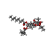
| Structure viewer | Molecule: MolmilJmol/JSmol |
|---|
- Downloads & links
-Download
| PDBx/mmCIF format | 1wpg.cif.gz | 784.6 KB | Display | PDBx/mmCIF format |
|---|---|---|---|---|
| PDB format | pdb1wpg.ent.gz | 639.1 KB | Display | PDB format |
| PDBx/mmJSON format | 1wpg.json.gz | Tree view | PDBx/mmJSON format | |
| Others |  Other downloads Other downloads |
-Validation report
| Arichive directory | https://data.pdbj.org/pub/pdb/validation_reports/wp/1wpgftp://data.pdbj.org/pub/pdb/validation_reports/wp/1wpg | HTTPS FTP |
|---|
-Related structure data
| Related structure data | 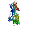 2zbdC  1fquS  1wpe S: Starting model for refinement C: citing same article ( |
|---|---|
| Similar structure data |


-Links
 PDBj
PDBj

- Assembly
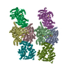
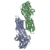
| Deposited unit | 
| ||||||||||||||||||||||||||||
|---|---|---|---|---|---|---|---|---|---|---|---|---|---|---|---|---|---|---|---|---|---|---|---|---|---|---|---|---|---|
| 1 |
| ||||||||||||||||||||||||||||
| Unit cell |
| ||||||||||||||||||||||||||||
| Noncrystallographic symmetry (NCS) | NCS oper:
|
-Components
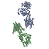
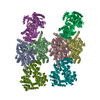
-Protein , 1 types, 4 molecules ABCD
| #1: Protein | Mass: 109602.578 Da / Num. of mol.: 4 / Source method: isolated from a natural source / Source: (natural) |
|---|
-Non-polymers , 6 types, 714 molecules 


| #2: Chemical | ChemComp-MG /  Mass: 24.305 Da / Num. of mol.: 8 / Source method: obtained synthetically / Formula: Mg Mass: 24.305 Da / Num. of mol.: 8 / Source method: obtained synthetically / Formula: Mg#3: Chemical | ChemComp-NA /  Mass: 22.990 Da / Num. of mol.: 4 / Source method: obtained synthetically / Formula: Na Mass: 22.990 Da / Num. of mol.: 4 / Source method: obtained synthetically / Formula: Na#4: Chemical | ChemComp-MF4 /  Mass: 100.299 Da / Num. of mol.: 4 / Source method: obtained synthetically / Formula: F4Mg Mass: 100.299 Da / Num. of mol.: 4 / Source method: obtained synthetically / Formula: F4Mg#5: Chemical | ChemComp-ADP /  Mass: 427.201 Da / Num. of mol.: 4 / Source method: obtained synthetically / Formula: C10H15N5O10P2 / Comment: ADP, energy-carrying molecule*YM Mass: 427.201 Da / Num. of mol.: 4 / Source method: obtained synthetically / Formula: C10H15N5O10P2 / Comment: ADP, energy-carrying molecule*YM#6: Chemical | ChemComp-TG1 /  Mass: 650.754 Da / Num. of mol.: 4 / Source method: obtained synthetically / Formula: C34H50O12 Mass: 650.754 Da / Num. of mol.: 4 / Source method: obtained synthetically / Formula: C34H50O12#7: Water | ChemComp-HOH / | Mass: 18.015 Da / Num. of mol.: 690 / Source method: isolated from a natural source / Formula: H2O |
|---|
-Details
| Has protein modification | Y |
|---|---|
| Sequence details | THE C-TERMINAL RESIDUES IN SWS ENTRY P04191 ARE FROM 994 TO 1001, DPEDERRK. IN ISOFORM SERCA1A, ...THE C-TERMINAL RESIDUES IN SWS ENTRY P04191 ARE FROM 994 TO 1001, DPEDERRK. IN ISOFORM SERCA1A, THERE IS ONLY ONE C-TERMINAL RESIDUE 994 G. |
-Experimental details
-Experiment
| Experiment | Method: X-RAY DIFFRACTION / Number of used crystals: 3 |
|---|
- Sample preparation
| Crystal | Density Matthews: 3.53 Å3/Da / Density % sol: 63.17 % |
|---|---|
| Crystal grow | Method: microdialysis / pH: 6.1 / Details: PEG400, pH 6.10, MICRODIALYSIS |
-Data collection
| Diffraction |
| |||||||||||||||
|---|---|---|---|---|---|---|---|---|---|---|---|---|---|---|---|---|
| Diffraction source |
| |||||||||||||||
| Detector |
| |||||||||||||||
| Radiation | Monochromator: ROTATED-INCLINED DOUBLE- CRYSTAL / Protocol: SINGLE WAVELENGTH / Monochromatic (M) / Laue (L): M / Scattering type: x-ray | |||||||||||||||
| Radiation wavelength |
| |||||||||||||||
| Reflection | Resolution: 2.3→20 Å / Num. obs: 250560 / % possible obs: 93.6 % / Observed criterion σ(I): -3 / Redundancy: 5.07 % / Biso Wilson estimate: 20.8 Å2 / Rmerge(I) obs: 0.059 / Net I/σ(I): 25.14 | |||||||||||||||
| Reflection shell | Resolution: 2.3→2.4 Å / Redundancy: 2.3 % / Mean I/σ(I) obs: 3.3 / Rsym value: 0.432 / % possible all: 81.9 |

- Processing
| Software |
| ||||||||||||||||||||||||||||||||||||
|---|---|---|---|---|---|---|---|---|---|---|---|---|---|---|---|---|---|---|---|---|---|---|---|---|---|---|---|---|---|---|---|---|---|---|---|---|---|
| Refinement | Method to determine structure: MOLECULAR REPLACEMENT Starting model: 3 CYTOPLASMIC DOMAINS OF CA2+-ATPASE IN 1FQU Resolution: 2.3→15 Å / Rfactor Rfree error: 0.002 / Data cutoff high absF: 410979.62 / Data cutoff low absF: 0 / Isotropic thermal model: RESTRAINED / Cross valid method: THROUGHOUT / σ(F): 2 / Stereochemistry target values: Engh & Huber
| ||||||||||||||||||||||||||||||||||||
| Solvent computation | Solvent model: FLAT MODEL / Bsol: 43.47 Å2 / ksol: 0.328798 e/Å3 | ||||||||||||||||||||||||||||||||||||
| Displacement parameters | Biso mean: 59.3 Å2
| ||||||||||||||||||||||||||||||||||||
| Refine analyze |
| ||||||||||||||||||||||||||||||||||||
| Refinement step | Cycle: LAST / Resolution: 2.3→15 Å
| ||||||||||||||||||||||||||||||||||||
| Refine LS restraints |
| ||||||||||||||||||||||||||||||||||||
| Refine LS restraints NCS | NCS model details: CONSTR | ||||||||||||||||||||||||||||||||||||
| LS refinement shell | Resolution: 2.3→2.44 Å / Rfactor Rfree error: 0.008 / Total num. of bins used: 6
| ||||||||||||||||||||||||||||||||||||
| Xplor file |
|
