 Movie
Movie Controller

[English] 日本語
 Yorodumi
- PDB-1mjg: CRYSTAL STRUCTURE OF BIFUNCTIONAL CARBON MONOXIDE DEHYDROGENASE/A... -
+ Open data

- Basic information
| Entry | Database: PDB / ID: 1mjg | ||||||
|---|---|---|---|---|---|---|---|
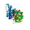
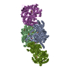
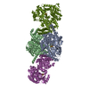
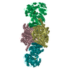
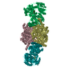
| Title | CRYSTAL STRUCTURE OF BIFUNCTIONAL CARBON MONOXIDE DEHYDROGENASE/ACETYL-COA SYNTHASE(CODH/ACS) FROM MOORELLA THERMOACETICA (F. CLOSTRIDIUM THERMOACETICUM) | ||||||
 Components Components |
| ||||||
 Keywords Keywords | OXIDOREDUCTASE / Carbon monoxide dehydrogenase(CODH) / Acetyl-CoA Synthase (ACS) / Nickel-Iron-Sulfur cluster (Ni-Fe-S) / Nickel-Coopper-Iron-Sulfur (Ni-Cu-Fe-S) Cluster / Hydrophobic CO channel / substrate tunnel / Helical domain / Rossmann fold / Electron transfer / Clostridium thermoaceticum / Wood-Ljundahl pathway | ||||||
| Function / homology |  Function and homology information Function and homology informationCO-methylating acetyl-CoA synthase / CO-methylating acetyl-CoA synthase activity / anaerobic carbon monoxide dehydrogenase / hydroxylamine reductase activity / anaerobic carbon-monoxide dehydrogenase activity / acetyl-CoA metabolic process / carbon fixation / nickel cation binding / peroxidase activity / generation of precursor metabolites and energy ...CO-methylating acetyl-CoA synthase / CO-methylating acetyl-CoA synthase activity / anaerobic carbon monoxide dehydrogenase / hydroxylamine reductase activity / anaerobic carbon-monoxide dehydrogenase activity / acetyl-CoA metabolic process / carbon fixation / nickel cation binding / peroxidase activity / generation of precursor metabolites and energy / response to hydrogen peroxide / 4 iron, 4 sulfur cluster binding / metal ion binding Similarity search - Function | ||||||
| Biological species |  Moorella thermoacetica (bacteria) Moorella thermoacetica (bacteria) | ||||||
| Method |  X-RAY DIFFRACTION / SYNCHROTRON / MAD, Molecular Replacement / Resolution: 2.2 Å X-RAY DIFFRACTION / SYNCHROTRON / MAD, Molecular Replacement / Resolution: 2.2 Å | ||||||
 Authors Authors | Doukov, T.I. / Iverson, T.M. / Seravalli, J. / Ragsdale, S.W. / Drennan, C.L. | ||||||
 Citation Citation | Journal: Science / Year: 2002 Title: A Ni-Fe-Cu center in a bifunctional carbon monoxide dehydrogenase/acetyl-CoA synthase Authors: Doukov, T.I. / Iverson, T.M. / Seravalli, J. / Ragsdale, S.W. / Drennan, C.L. | ||||||
| History |
| ||||||
| Remark 600 | HETEROGEN The A-Cluster consists of FS4 900, CU 901 and NI 902 for chains M,N,O,P. There is ...HETEROGEN The A-Cluster consists of FS4 900, CU 901 and NI 902 for chains M,N,O,P. There is electron density for a ligand on the Cu of the A-cluster. An acetyl moiety (ACT) has been refined in this position, although the exact nature of this ligand is unknown. | ||||||
| Remark 999 | SEQUENCE Position 685 of the ACS subunit (chains M,N,O,P) has been modeled as Ser according to the ...SEQUENCE Position 685 of the ACS subunit (chains M,N,O,P) has been modeled as Ser according to the PIR B41670 sequence entry. The Swissprot P27988 entry lists position 685 as Arg. The electron density can be interpreted as Arg or as Ser hydrogen bonded to water molecules. |
- Structure visualization
| Structure viewer | Molecule: MolmilJmol/JSmol |
|---|
- Downloads & links
-Download
| PDBx/mmCIF format | 1mjg.cif.gz | 1 MB | Display | PDBx/mmCIF format |
|---|---|---|---|---|
| PDB format | pdb1mjg.ent.gz | 883.5 KB | Display | PDB format |
| PDBx/mmJSON format | 1mjg.json.gz | Tree view | PDBx/mmJSON format | |
| Others |  Other downloads Other downloads |
-Validation report
| Arichive directory | https://data.pdbj.org/pub/pdb/validation_reports/mj/1mjgftp://data.pdbj.org/pub/pdb/validation_reports/mj/1mjg | HTTPS FTP |
|---|
-Related structure data
| Related structure data | 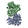 1jqkS S: Starting model for refinement |
|---|---|
| Similar structure data |


-Links
 PDBj
PDBj- Assembly
| Deposited unit | 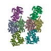
| ||||||||
|---|---|---|---|---|---|---|---|---|---|
| 1 | 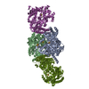
| ||||||||
| 2 | 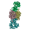
| ||||||||
| Unit cell |
|
-Components
-Protein , 2 types, 8 molecules ABCDMNOP
| #1: Protein | Mass: 73006.930 Da / Num. of mol.: 4 / Source method: isolated from a natural source / Source: (natural) Moorella thermoacetica (bacteria)References: UniProt: P27989, carbon-monoxide dehydrogenase (acceptor) #2: Protein | Mass: 81746.812 Da / Num. of mol.: 4 / Source method: isolated from a natural source / Source: (natural) Moorella thermoacetica (bacteria)References: UniProt: P27988, carbon-monoxide dehydrogenase (acceptor) |
|---|
-Non-polymers , 6 types, 1255 molecules 


| #3: Chemical | ChemComp-SF4 /  Mass: 351.640 Da / Num. of mol.: 10 / Source method: obtained synthetically / Formula: Fe4S4 Mass: 351.640 Da / Num. of mol.: 10 / Source method: obtained synthetically / Formula: Fe4S4#4: Chemical | ChemComp-XCC /  Mass: 410.333 Da / Num. of mol.: 4 / Source method: obtained synthetically / Formula: Fe4NiS4 Mass: 410.333 Da / Num. of mol.: 4 / Source method: obtained synthetically / Formula: Fe4NiS4#5: Chemical | ChemComp-CU1 /  Mass: 63.546 Da / Num. of mol.: 4 / Source method: obtained synthetically / Formula: Cu Mass: 63.546 Da / Num. of mol.: 4 / Source method: obtained synthetically / Formula: Cu#6: Chemical | ChemComp-NI /  Mass: 58.693 Da / Num. of mol.: 4 / Source method: obtained synthetically / Formula: Ni Mass: 58.693 Da / Num. of mol.: 4 / Source method: obtained synthetically / Formula: Ni#7: Chemical | ChemComp-ACT /  Mass: 59.044 Da / Num. of mol.: 4 / Source method: obtained synthetically / Formula: C2H3O2 Mass: 59.044 Da / Num. of mol.: 4 / Source method: obtained synthetically / Formula: C2H3O2#8: Water | ChemComp-HOH / | Mass: 18.015 Da / Num. of mol.: 1229 / Source method: isolated from a natural source / Formula: H2O |
|---|
-Details
| Has protein modification | Y |
|---|
-Experimental details
-Experiment
| Experiment | Method: X-RAY DIFFRACTION / Number of used crystals: 1 |
|---|
- Sample preparation
| Crystal | Density Matthews: 2.73 Å3/Da / Density % sol: 54.96 % | ||||||||||||||||||||||||||||||||||||||||||||||||
|---|---|---|---|---|---|---|---|---|---|---|---|---|---|---|---|---|---|---|---|---|---|---|---|---|---|---|---|---|---|---|---|---|---|---|---|---|---|---|---|---|---|---|---|---|---|---|---|---|---|
| Crystal grow | Temperature: 298 K / Method: vapor diffusion, sitting drop / pH: 6.5 Details: 8 % PEG monomethyl ether 5000, 20% Glycerol, 200 mM Calcium Acetate, 100 mM PIPES pH 6.5, 2 mM dithioerythritol, VAPOR DIFFUSION, SITTING DROP, temperature 298.0K | ||||||||||||||||||||||||||||||||||||||||||||||||
| Crystal grow | *PLUS pH: 7.6 | ||||||||||||||||||||||||||||||||||||||||||||||||
| Components of the solutions | *PLUS
|
-Data collection
| Diffraction | Mean temperature: 100 K |
|---|---|
| Diffraction source | Source: SYNCHROTRON / Site: ALS  / Beamline: 5.0.2 / Wavelength: 1.2 Å / Beamline: 5.0.2 / Wavelength: 1.2 Å |
| Detector | Type: ADSC QUANTUM 4 / Detector: CCD |
| Radiation | Protocol: SINGLE WAVELENGTH / Monochromatic (M) / Laue (L): M / Scattering type: x-ray |
| Radiation wavelength | Wavelength: 1.2 Å / Relative weight: 1 |
| Reflection | Resolution: 2.2→50 Å / Num. all: 332287 / Num. obs: 313013 / % possible obs: 95.1 % / Observed criterion σ(F): 0 / Observed criterion σ(I): -1 / Redundancy: 3.3 % / Biso Wilson estimate: 30.4 Å2 / Rsym value: 0.093 / Net I/σ(I): 12 |
| Reflection shell | Resolution: 2.2→2.28 Å / Rsym value: 0.39 / % possible all: 91.3 |
| Reflection | *PLUS Lowest resolution: 50 Å / Num. measured all: 1031478 / Rmerge(I) obs: 0.093 |
| Reflection shell | *PLUS % possible obs: 91.3 % / Rmerge(I) obs: 0.39 |
- Processing
| Software |
| ||||||||||||||||||||||||||||||||||||||||||||||||||||||||||||||||||||||||||||||||
|---|---|---|---|---|---|---|---|---|---|---|---|---|---|---|---|---|---|---|---|---|---|---|---|---|---|---|---|---|---|---|---|---|---|---|---|---|---|---|---|---|---|---|---|---|---|---|---|---|---|---|---|---|---|---|---|---|---|---|---|---|---|---|---|---|---|---|---|---|---|---|---|---|---|---|---|---|---|---|---|---|---|
| Refinement | Method to determine structure: MAD, Molecular Replacement Starting model: PDB ENTRY 1JQK Resolution: 2.2→20 Å / Isotropic thermal model: anisotropic / Cross valid method: THROUGHOUT / σ(F): 0 / Stereochemistry target values: Engh & Huber Details: Disordered regions include residues 1-2 and 536-540 of the CODH subunits (chains A-D) and residues 1, 313-320 of the ACS subunits (chains M-P). Also, the second and third domains of ACS ...Details: Disordered regions include residues 1-2 and 536-540 of the CODH subunits (chains A-D) and residues 1, 313-320 of the ACS subunits (chains M-P). Also, the second and third domains of ACS subunit chain O have poor electron density and high B factors compared to the other ACS subunits.
| ||||||||||||||||||||||||||||||||||||||||||||||||||||||||||||||||||||||||||||||||
| Solvent computation | Solvent model: CNS | ||||||||||||||||||||||||||||||||||||||||||||||||||||||||||||||||||||||||||||||||
| Displacement parameters | Biso mean: 41 Å2
| ||||||||||||||||||||||||||||||||||||||||||||||||||||||||||||||||||||||||||||||||
| Refinement step | Cycle: LAST / Resolution: 2.2→20 Å
| ||||||||||||||||||||||||||||||||||||||||||||||||||||||||||||||||||||||||||||||||
| Refine LS restraints |
| ||||||||||||||||||||||||||||||||||||||||||||||||||||||||||||||||||||||||||||||||
| Refine LS restraints NCS | NCS model details: RESTRAINTS | ||||||||||||||||||||||||||||||||||||||||||||||||||||||||||||||||||||||||||||||||
| LS refinement shell | Resolution: 2.2→2.3 Å / Total num. of bins used: 8 /
| ||||||||||||||||||||||||||||||||||||||||||||||||||||||||||||||||||||||||||||||||
| Xplor file | Serial no: 1 / Param file: PROTEIN_REP.PARAM / Topol file: PROTEIN.TOP | ||||||||||||||||||||||||||||||||||||||||||||||||||||||||||||||||||||||||||||||||
| Refinement | *PLUS Lowest resolution: 20 Å / Rfactor Rfree: 0.252 / Rfactor Rwork: 0.22 | ||||||||||||||||||||||||||||||||||||||||||||||||||||||||||||||||||||||||||||||||
| Solvent computation | *PLUS | ||||||||||||||||||||||||||||||||||||||||||||||||||||||||||||||||||||||||||||||||
| Displacement parameters | *PLUS | ||||||||||||||||||||||||||||||||||||||||||||||||||||||||||||||||||||||||||||||||
| Refine LS restraints | *PLUS
|
