 Movie
Movie Controller

[English] 日本語
 Yorodumi
- PDB-1e2u: Low Temperature Structure of Hybrid Cluster Protein from Desulfov... -
+ Open data

- Basic information
| Entry | Database: PDB / ID: 1e2u | ||||||
|---|---|---|---|---|---|---|---|
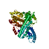
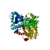
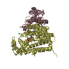
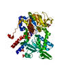
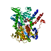
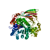
| Title | Low Temperature Structure of Hybrid Cluster Protein from Desulfovibrio vulgaris to 1.6A | ||||||
 Components Components | HYDROXYLAMINE REDUCTASE | ||||||
 Keywords Keywords | OXIDOREDUCTASE / CUBANE / IRON SULFUR / FUSCOREDOXIN | ||||||
| Function / homology |  Function and homology information Function and homology informationhydroxylamine reductase / hydroxylamine reductase activity / peroxidase activity / response to hydrogen peroxide / 4 iron, 4 sulfur cluster binding / metal ion binding / cytoplasm Similarity search - Function | ||||||
| Biological species |  DESULFOVIBRIO VULGARIS (bacteria) DESULFOVIBRIO VULGARIS (bacteria) | ||||||
| Method |  X-RAY DIFFRACTION / SYNCHROTRON / MOLECULAR REPLACEMENT / Resolution: 1.6 Å X-RAY DIFFRACTION / SYNCHROTRON / MOLECULAR REPLACEMENT / Resolution: 1.6 Å | ||||||
 Authors Authors | Cooper, S.J. / Bailey, S. / Hagen, W.R. / Lindley, P.F. | ||||||
 Citation Citation | Journal: Biochemistry / Year: 2000 Title: Hybrid-Cluster Protein (Hcp) from Desulfovibrio Vulgaris (Hildenborough) at 1.6 A Resolution. Authors: Cooper, S.J. / Garner, C.D. / Hagen, W.R. / Lindley, P.F. / Bailey, S. | ||||||
| History |
|
- Structure visualization
| Structure viewer | Molecule: MolmilJmol/JSmol |
|---|
- Downloads & links
-Download
| PDBx/mmCIF format | 1e2u.cif.gz | 132 KB | Display | PDBx/mmCIF format |
|---|---|---|---|---|
| PDB format | pdb1e2u.ent.gz | 101.6 KB | Display | PDB format |
| PDBx/mmJSON format | 1e2u.json.gz | Tree view | PDBx/mmJSON format | |
| Others |  Other downloads Other downloads |
-Validation report
| Arichive directory | https://data.pdbj.org/pub/pdb/validation_reports/e2/1e2uftp://data.pdbj.org/pub/pdb/validation_reports/e2/1e2u | HTTPS FTP |
|---|
-Related structure data
| Related structure data |  1e1dSC S: Starting model for refinement C: citing same article ( |
|---|---|
| Similar structure data |


-Links
 PDBj
PDBj- Assembly
| Deposited unit | 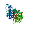
| ||||||||
|---|---|---|---|---|---|---|---|---|---|
| 1 |
| ||||||||
| Unit cell |
|
-Components
| #1: Protein | Mass: 60074.703 Da / Num. of mol.: 1 Source method: isolated from a genetically manipulated source Source: (gene. exp.) DESULFOVIBRIO VULGARIS (bacteria) / Strain: HILDENBOROUGH NCIMB8303 / Plasmid: PJSP104 / Production host: DESULFOVIBRIO VULGARIS (bacteria) / Strain (production host): HILDENBOROUGH NCIMB8303 / References: UniProt: P31101 |
|---|---|
| #2: Chemical | ChemComp-SF4 /   Mass: 351.640 Da / Num. of mol.: 1 / Source method: obtained synthetically / Formula: Fe4S4 Mass: 351.640 Da / Num. of mol.: 1 / Source method: obtained synthetically / Formula: Fe4S4 |
| #3: Chemical | ChemComp-FSO /   Mass: 367.573 Da / Num. of mol.: 1 / Source method: obtained synthetically / Formula: Fe4O3S3 Mass: 367.573 Da / Num. of mol.: 1 / Source method: obtained synthetically / Formula: Fe4O3S3 |
| #4: Water | ChemComp-HOH /  Mass: 18.015 Da / Num. of mol.: 630 / Source method: isolated from a natural source / Formula: H2O Mass: 18.015 Da / Num. of mol.: 630 / Source method: isolated from a natural source / Formula: H2O |
| Has protein modification | Y |
| Nonpolymer details | HET GROUP: CSS CSS-S-MERCAPTOCYSTEINE LIGAND TO HYBRID CLUSTER HET GROUP: FS4 FS4-CUBANE IRON ...HET GROUP: CSS CSS-S-MERCAPTOCY |
| Sequence details | MODRES: 1E2U CSS A 406() THIOCYSTEI |
-Experimental details
-Experiment
| Experiment | Method: X-RAY DIFFRACTION / Number of used crystals: 1 |
|---|
- Sample preparation
| Crystal | Density Matthews: 2.6 Å3/Da / Density % sol: 52.5 % | ||||||||||||||||||||||||||||||||||||
|---|---|---|---|---|---|---|---|---|---|---|---|---|---|---|---|---|---|---|---|---|---|---|---|---|---|---|---|---|---|---|---|---|---|---|---|---|---|
| Crystal grow | pH: 5.9 Details: PROTEIN WAS CRYSTALLISED FROM 0.1M MES PH5.9, 60MM MAGNESIUM ACETATE AND 25-30% PEG 8000 | ||||||||||||||||||||||||||||||||||||
| Crystal grow | *PLUS Temperature: 277 K / Method: vapor diffusion, hanging dropDetails: drop consists of equal amounts of protein and precipitant solutions | ||||||||||||||||||||||||||||||||||||
| Components of the solutions | *PLUS
|
-Data collection
| Diffraction | Mean temperature: 100 K |
|---|---|
| Diffraction source | Source: SYNCHROTRON / Site: SRS  / Beamline: PX7.2 / Wavelength: 1.488 / Beamline: PX7.2 / Wavelength: 1.488 |
| Detector | Type: MARRESEARCH / Detector: IMAGE PLATE / Date: Jun 15, 1998 |
| Radiation | Protocol: SINGLE WAVELENGTH / Monochromatic (M) / Laue (L): M / Scattering type: x-ray |
| Radiation wavelength | Wavelength: 1.488 Å / Relative weight: 1 |
| Reflection | Resolution: 1.59→20 Å / Num. obs: 77446 / % possible obs: 91.3 % / Redundancy: 2.5 % / Biso Wilson estimate: 11.436 Å2 / Rsym value: 0.069 / Net I/σ(I): 5.6 |
| Reflection shell | Resolution: 1.59→1.68 Å / Redundancy: 2.4 % / Mean I/σ(I) obs: 6.5 / Rsym value: 0.1 / % possible all: 87.7 |
| Reflection | *PLUS Rmerge(I) obs: 0.07 |
| Reflection shell | *PLUS % possible obs: 87.7 % / Rmerge(I) obs: 0.1 |
- Processing
| Software |
| ||||||||||||||||||||||||||||||||||||||||||||||||||||||||||||||||||||||||||||||||||||
|---|---|---|---|---|---|---|---|---|---|---|---|---|---|---|---|---|---|---|---|---|---|---|---|---|---|---|---|---|---|---|---|---|---|---|---|---|---|---|---|---|---|---|---|---|---|---|---|---|---|---|---|---|---|---|---|---|---|---|---|---|---|---|---|---|---|---|---|---|---|---|---|---|---|---|---|---|---|---|---|---|---|---|---|---|---|
| Refinement | Method to determine structure: MOLECULAR REPLACEMENT Starting model: PDB ENTRY 1E1D Resolution: 1.6→20 Å / SU B: 1.26981 / SU ML: 0.04585 / Cross valid method: THROUGHOUT / σ(F): 0 / ESU R: 0.08154 / ESU R Free: 0.08003 / Details: METAL CLUSTERS REFINED WITH ANISOTROPIC B
| ||||||||||||||||||||||||||||||||||||||||||||||||||||||||||||||||||||||||||||||||||||
| Displacement parameters | Biso mean: 13.443 Å2 | ||||||||||||||||||||||||||||||||||||||||||||||||||||||||||||||||||||||||||||||||||||
| Refinement step | Cycle: LAST / Resolution: 1.6→20 Å
| ||||||||||||||||||||||||||||||||||||||||||||||||||||||||||||||||||||||||||||||||||||
| Refine LS restraints |
| ||||||||||||||||||||||||||||||||||||||||||||||||||||||||||||||||||||||||||||||||||||
| Software | *PLUS Name: REFMAC / Classification: refinement | ||||||||||||||||||||||||||||||||||||||||||||||||||||||||||||||||||||||||||||||||||||
| Refinement | *PLUS Rfactor Rfree: 0.183 | ||||||||||||||||||||||||||||||||||||||||||||||||||||||||||||||||||||||||||||||||||||
| Solvent computation | *PLUS | ||||||||||||||||||||||||||||||||||||||||||||||||||||||||||||||||||||||||||||||||||||
| Displacement parameters | *PLUS |
