Resolution: 2.9→2.97 Å / Redundancy: 5.7 % / Rmerge(I) obs: 0.8 / Mean I/σ(I) obs: 2.1 / Num. unique all: 2554 / Rsym value: 0.8 / % possible all: 99.8
-
Processing
Software
Name
Version
Classification
REFMAC
5.1.9999
refinement
DENZO
datareduction
SCALEPACK
datascaling
CNS
phasing
Refinement
Method to determine structure: MOLECULAR REPLACEMENT / Resolution: 2.9→50 Å / Cor.coef. Fo:Fc: 0.922 / Cor.coef. Fo:Fc free: 0.872 / SU B: 33.012 / SU ML: 0.292 / TLS residual ADP flag: LIKELY RESIDUAL / Cross valid method: THROUGHOUT / σ(F): 0 / ESU R: 0.774 / ESU R Free: 0.39 / Stereochemistry target values: Engh & Huber Details: Hydrogens have been added in the riding positions. Identification of a potassium ion binding site was verified by difference Fourier maps using a data set collected on a crystal prepared ...Details: Hydrogens have been added in the riding positions. Identification of a potassium ion binding site was verified by difference Fourier maps using a data set collected on a crystal prepared under conditions with rubidium substituting for potassium. This additional data set is also included in the accompanying structure factor file.
Rfactor
Num. reflection
% reflection
Selection details
Rfree
0.29348
1153
3 %
RANDOM
Rwork
0.24602
-
-
-
all
0.24746
38895
-
-
obs
0.24746
37727
99.88 %
-
Solvent computation
Ion probe radii: 0.8 Å / Shrinkage radii: 0.8 Å / VDW probe radii: 1.2 Å / Solvent model: BABINET MODEL WITH MASK
Displacement parameters
Biso mean: 74.729 Å2
Baniso -1
Baniso -2
Baniso -3
1-
-1.34 Å2
0 Å2
-3.83 Å2
2-
-
6.38 Å2
0 Å2
3-
-
-
-7.51 Å2
Refinement step
Cycle: LAST / Resolution: 2.9→50 Å
Protein
Nucleic acid
Ligand
Solvent
Total
Num. atoms
7671
0
37
43
7751
Refine LS restraints
Refine-ID
Type
Dev ideal
Dev ideal target
Number
X-RAY DIFFRACTION
r_bond_refined_d
0.01
0.022
7845
X-RAY DIFFRACTION
r_bond_other_d
0.001
0.02
7322
X-RAY DIFFRACTION
r_angle_refined_deg
1.571
1.963
10645
X-RAY DIFFRACTION
r_angle_other_deg
0.878
3
17030
X-RAY DIFFRACTION
r_dihedral_angle_1_deg
7.662
5
993
X-RAY DIFFRACTION
r_dihedral_angle_2_deg
35.537
24.357
319
X-RAY DIFFRACTION
r_dihedral_angle_3_deg
19.904
15
1382
X-RAY DIFFRACTION
r_dihedral_angle_4_deg
19.061
15
48
X-RAY DIFFRACTION
r_chiral_restr
0.085
0.2
1239
X-RAY DIFFRACTION
r_gen_planes_refined
0.004
0.02
8638
X-RAY DIFFRACTION
r_gen_planes_other
0.001
0.02
1508
X-RAY DIFFRACTION
r_nbd_refined
0.251
0.2
2187
X-RAY DIFFRACTION
r_nbd_other
0.203
0.2
8027
X-RAY DIFFRACTION
r_nbtor_refined
X-RAY DIFFRACTION
r_nbtor_other
0.093
0.2
4689
X-RAY DIFFRACTION
r_xyhbond_nbd_refined
0.195
0.2
249
X-RAY DIFFRACTION
r_xyhbond_nbd_other
X-RAY DIFFRACTION
r_metal_ion_refined
0.156
0.2
12
X-RAY DIFFRACTION
r_metal_ion_other
X-RAY DIFFRACTION
r_symmetry_vdw_refined
0.157
0.2
7
X-RAY DIFFRACTION
r_symmetry_vdw_other
0.23
0.2
31
X-RAY DIFFRACTION
r_symmetry_hbond_refined
0.175
0.2
5
X-RAY DIFFRACTION
r_symmetry_hbond_other
X-RAY DIFFRACTION
r_mcbond_it
0.607
1.5
4947
X-RAY DIFFRACTION
r_mcbond_other
0.074
1.5
2013
X-RAY DIFFRACTION
r_mcangle_it
1.139
2
8012
X-RAY DIFFRACTION
r_scbond_it
1.353
3
2921
X-RAY DIFFRACTION
r_scangle_it
2.334
4.5
2633
X-RAY DIFFRACTION
r_rigid_bond_restr
X-RAY DIFFRACTION
r_sphericity_free
X-RAY DIFFRACTION
r_sphericity_bonded
LS refinement shell
Resolution: 2.9→2.975 Å / Total num. of bins used: 20 /
Rfactor
Num. reflection
Rfree
0.405
73
Rwork
0.335
2763
Refinement TLS params.
Method: refined / Refine-ID: X-RAY DIFFRACTION
ID
L11 (°2)
L12 (°2)
L13 (°2)
L22 (°2)
L23 (°2)
L33 (°2)
S11 (Å °)
S12 (Å °)
S13 (Å °)
S21 (Å °)
S22 (Å °)
S23 (Å °)
S31 (Å °)
S32 (Å °)
S33 (Å °)
T11 (Å2)
T12 (Å2)
T13 (Å2)
T22 (Å2)
T23 (Å2)
T33 (Å2)
Origin x (Å)
Origin y (Å)
Origin z (Å)
1
0.8551
-0.0813
0.2487
0.6936
-0.481
1.5339
0.1417
-0.0274
-0.4248
-0.0207
0.1403
-0.0203
0.4036
0.2088
-0.282
-0.1983
0.0123
-0.0076
0.2055
-0.0326
-0.3813
-6.5328
-9.2965
64.1621
2
0.5447
0.0618
0.6097
0.6412
-0.1545
3.1441
-0.1114
-0.2164
0.0799
0.0871
0.0297
-0.0906
-0.4352
0.3302
0.0817
-0.1419
-0.1151
-0.0191
0.2883
-0.1271
-0.5324
-5.5108
9.742
71.2839
3
1.4342
0.1785
0.2509
0.3905
-0.1567
1.658
0.2395
-0.0932
-0.1092
-0.0722
-0.2307
0.0559
-0.0545
-0.1317
-0.0088
-0.0962
0.024
-0.0121
-0.163
-0.0259
0.0216
-21.8451
-4.5035
11.6936
Refinement TLS group
ID
Refine-ID
Refine TLS-ID
Auth asym-ID
Label asym-ID
Auth seq-ID
Label seq-ID
1
X-RAY DIFFRACTION
1
A
A
45 - 122
45 - 122
2
X-RAY DIFFRACTION
1
A
A
238 - 329
238 - 329
3
X-RAY DIFFRACTION
2
A
A
742 - 994
742 - 994
4
X-RAY DIFFRACTION
2
A
C - D
1003 - 1004
1
5
X-RAY DIFFRACTION
3
A
A
1 - 44
1 - 44
6
X-RAY DIFFRACTION
3
A
A
123 - 237
123 - 237
7
X-RAY DIFFRACTION
3
A
A
330 - 741
330 - 741
8
X-RAY DIFFRACTION
3
A
H - B
1001 - 1002
1
9
X-RAY DIFFRACTION
3
A
E - G
1005 - 1007
1
+
About Yorodumi
-
News
-
Feb 9, 2022. New format data for meta-information of EMDB entries
New format data for meta-information of EMDB entries
Version 3 of the EMDB header file is now the official format.
The previous official version 1.9 will be removed from the archive.
In the structure databanks used in Yorodumi, some data are registered as the other names, "COVID-19 virus" and "2019-nCoV". Here are the details of the virus and the list of structure data.
Jan 31, 2019. EMDB accession codes are about to change! (news from PDBe EMDB page)
EMDB accession codes are about to change! (news from PDBe EMDB page)
The allocation of 4 digits for EMDB accession codes will soon come to an end. Whilst these codes will remain in use, new EMDB accession codes will include an additional digit and will expand incrementally as the available range of codes is exhausted. The current 4-digit format prefixed with “EMD-” (i.e. EMD-XXXX) will advance to a 5-digit format (i.e. EMD-XXXXX), and so on. It is currently estimated that the 4-digit codes will be depleted around Spring 2019, at which point the 5-digit format will come into force.
The EM Navigator/Yorodumi systems omit the EMD- prefix.
Related info.:Q: What is EMD? / ID/Accession-code notation in Yorodumi/EM Navigator
Yorodumi is a browser for structure data from EMDB, PDB, SASBDB, etc.
This page is also the successor to EM Navigator detail page, and also detail information page/front-end page for Omokage search.
The word "yorodu" (or yorozu) is an old Japanese word meaning "ten thousand". "mi" (miru) is to see.
Related info.:EMDB / PDB / SASBDB / Comparison of 3 databanks / Yorodumi Search / Aug 31, 2016. New EM Navigator & Yorodumi / Yorodumi Papers / Jmol/JSmol / Function and homology information / Changes in new EM Navigator and Yorodumi
 Movie
Movie Controller


 Open data
 Basic information
 Components
 Keywords
 Function and homology information
 X-RAY DIFFRACTION /
  Authors
 Citation
 Structure visualization
 Downloads & links
 Other downloads
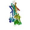


 PDBj
PDBj


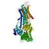
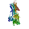
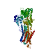
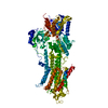
 Assembly


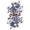
 Mass: 102.975 Da / Num. of mol.: 1 / Source method: obtained synthetically / Formula: AlF4
 Mass: 40.078 Da / Num. of mol.: 2 / Source method: obtained synthetically / Formula: Ca
 Mass: 24.305 Da / Num. of mol.: 2 / Source method: obtained synthetically / Formula: Mg
 Mass: 39.098 Da / Num. of mol.: 1 / Source method: obtained synthetically / Formula: K
 Mass: 427.201 Da / Num. of mol.: 1 / Source method: obtained synthetically / Formula: C10H15N5O10P2 / Comment: ADP, energy-carrying molecule*YM
 Sample preparation
 Processing