 Movie
Movie Controller


+ Open data

- Basic information
| Entry | Database: PDB / ID: 2za4 | ||||||
|---|---|---|---|---|---|---|---|
| Title | Crystal Structural Analysis of Barnase-barstar Complex | ||||||
 Components Components |
| ||||||
 Keywords Keywords | Hydrolase/Hydrolase Inhibitor / protein-protein complex / Endonuclease / Genetically modified food / Hydrolase / Nuclease / Secreted / Cytoplasm / Hydrolase-Hydrolase Inhibitor COMPLEX | ||||||
| Function / homology |  Function and homology information Function and homology informationHydrolases; Acting on ester bonds; Endoribonucleases producing 3'-phosphomonoesters / RNA endonuclease activity / hydrolase activity / RNA binding / extracellular region / cytoplasm Similarity search - Function | ||||||
| Biological species |  | ||||||
| Method |  X-RAY DIFFRACTION / SYNCHROTRON / MOLECULAR REPLACEMENT / Resolution: 1.58 Å X-RAY DIFFRACTION / SYNCHROTRON / MOLECULAR REPLACEMENT / Resolution: 1.58 Å | ||||||
 Authors Authors | Urakubo, Y. / Ikura, T. / Ito, N. | ||||||
 Citation Citation | Journal: Protein Sci. / Year: 2008 Title: Crystal structural analysis of protein-protein interactions drastically destabilized by a single mutation Authors: Urakubo, Y. / Ikura, T. / Ito, N. | ||||||
| History |
|
- Structure visualization
| Structure viewer | Molecule: MolmilJmol/JSmol |
|---|
- Downloads & links
-Download
| PDBx/mmCIF format | 2za4.cif.gz | 99.4 KB | Display | PDBx/mmCIF format |
|---|---|---|---|---|
| PDB format | pdb2za4.ent.gz | 75 KB | Display | PDB format |
| PDBx/mmJSON format | 2za4.json.gz | Tree view | PDBx/mmJSON format | |
| Others |  Other downloads Other downloads |
-Validation report
| Arichive directory | https://data.pdbj.org/pub/pdb/validation_reports/za/2za4ftp://data.pdbj.org/pub/pdb/validation_reports/za/2za4 | HTTPS FTP |
|---|
-Related structure data
| Related structure data |  1brsS S: Starting model for refinement |
|---|---|
| Similar structure data |


-Links
 PDBj
PDBj- Assembly
| Deposited unit | 
| ||||||||
|---|---|---|---|---|---|---|---|---|---|
| 1 | 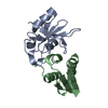
| ||||||||
| 2 | 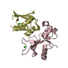
| ||||||||
| 3 | 
| ||||||||
| 4 | 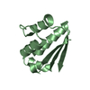
| ||||||||
| 5 | 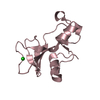
| ||||||||
| 6 | 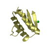
| ||||||||
| Unit cell |
|
-Components
| #1: Protein | Mass: 12340.618 Da / Num. of mol.: 2 / Mutation: K98A Source method: isolated from a genetically manipulated source Source: (gene. exp.) References: UniProt: P00648, Hydrolases; Acting on ester bonds; Endoribonucleases producing 3'-phosphomonoesters #2: Protein | Mass: 10244.598 Da / Num. of mol.: 2 / Mutation: D39A, C40A, C82A Source method: isolated from a genetically manipulated source Source: (gene. exp.) #3: Chemical |   Mass: 35.453 Da / Num. of mol.: 2 / Source method: obtained synthetically / Formula: Cl Mass: 35.453 Da / Num. of mol.: 2 / Source method: obtained synthetically / Formula: Cl#4: Water | ChemComp-HOH / |  Mass: 18.015 Da / Num. of mol.: 448 / Source method: isolated from a natural source / Formula: H2O Mass: 18.015 Da / Num. of mol.: 448 / Source method: isolated from a natural source / Formula: H2O |
|---|
-Experimental details
-Experiment
| Experiment | Method: X-RAY DIFFRACTION / Number of used crystals: 1 |
|---|
- Sample preparation
| Crystal | Density Matthews: 2.55 Å3/Da / Density % sol: 51.7 % |
|---|---|
| Crystal grow | Temperature: 293 K / Method: vapor diffusion, hanging drop / pH: 7 Details: 20% PEG 6000, 0.1M HEPES (pH 7.0), 1.0M lithium chloride, VAPOR DIFFUSION, HANGING DROP, temperature 293K |
-Data collection
| Diffraction | Mean temperature: 95 K |
|---|---|
| Diffraction source | Source: SYNCHROTRON / Site: Photon Factory  / Beamline: BL-6A / Wavelength: 0.978 Å / Beamline: BL-6A / Wavelength: 0.978 Å |
| Detector | Type: ADSC QUANTUM 4 / Detector: CCD / Date: Jan 29, 2006 |
| Radiation | Protocol: SINGLE WAVELENGTH / Monochromatic (M) / Laue (L): M / Scattering type: x-ray |
| Radiation wavelength | Wavelength: 0.978 Å / Relative weight: 1 |
| Reflection | Resolution: 1.58→50 Å / Num. all: 61328 / Num. obs: 61328 / % possible obs: 99.3 % / Observed criterion σ(F): 0 / Observed criterion σ(I): 0 / Redundancy: 3.1 % / Biso Wilson estimate: 19.3 Å2 / Rmerge(I) obs: 0.047 / Rsym value: 0.047 / Net I/σ(I): 35.1 |
| Reflection shell | Resolution: 1.58→1.61 Å / Redundancy: 2.7 % / Rmerge(I) obs: 0.246 / Mean I/σ(I) obs: 5 / Num. unique all: 3459 / Rsym value: 0.246 / % possible all: 90.9 |
- Processing
| Software |
| ||||||||||||||||||||||||||||||||||||||||||||||||||||||||||||
|---|---|---|---|---|---|---|---|---|---|---|---|---|---|---|---|---|---|---|---|---|---|---|---|---|---|---|---|---|---|---|---|---|---|---|---|---|---|---|---|---|---|---|---|---|---|---|---|---|---|---|---|---|---|---|---|---|---|---|---|---|---|
| Refinement | Method to determine structure: MOLECULAR REPLACEMENT Starting model: PDB ENTRY 1BRS Resolution: 1.58→43.36 Å / Rfactor Rfree error: 0.004 / Data cutoff high absF: 143527.46 / Data cutoff low absF: 0 / Isotropic thermal model: RESTRAINED / Cross valid method: THROUGHOUT / σ(F): 0 / σ(I): 0
| ||||||||||||||||||||||||||||||||||||||||||||||||||||||||||||
| Solvent computation | Solvent model: FLAT MODEL / Bsol: 53.2632 Å2 / ksol: 0.369678 e/Å3 | ||||||||||||||||||||||||||||||||||||||||||||||||||||||||||||
| Displacement parameters | Biso mean: 22.4 Å2
| ||||||||||||||||||||||||||||||||||||||||||||||||||||||||||||
| Refine analyze |
| ||||||||||||||||||||||||||||||||||||||||||||||||||||||||||||
| Refinement step | Cycle: LAST / Resolution: 1.58→43.36 Å
| ||||||||||||||||||||||||||||||||||||||||||||||||||||||||||||
| Refine LS restraints |
| ||||||||||||||||||||||||||||||||||||||||||||||||||||||||||||
| LS refinement shell | Resolution: 1.58→1.68 Å / Rfactor Rfree error: 0.014 / Total num. of bins used: 6
| ||||||||||||||||||||||||||||||||||||||||||||||||||||||||||||
| Xplor file |
|
