 Movie
Movie Controller


+ Open data

- Basic information
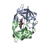
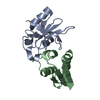
| Entry | Database: PDB / ID: 1b2u | ||||||
|---|---|---|---|---|---|---|---|
| Title | STRUCTURAL RESPONSE TO MUTATION AT A PROTEIN-PROTEIN INTERFACE | ||||||
 Components Components |
| ||||||
 Keywords Keywords | HYDROLASE/HYDROLASE INHIBITOR / RNASE-INHIBITOR COMPLEX / INTERFACIAL DOUBLE MUTANT / HYDROLASE-HYDROLASE INHIBITOR COMPLEX | ||||||
| Function / homology |  Function and homology information Function and homology informationHydrolases; Acting on ester bonds; Endoribonucleases producing 3'-phosphomonoesters / RNA endonuclease activity / hydrolase activity / RNA binding / extracellular region / cytoplasm Similarity search - Function | ||||||
| Biological species |  | ||||||
| Method |  X-RAY DIFFRACTION / OTHER / Resolution: 2.1 Å X-RAY DIFFRACTION / OTHER / Resolution: 2.1 Å | ||||||
 Authors Authors | Vaughan, C.K. / Buckle, A.M. / Fersht, A.R. | ||||||
 Citation Citation | Journal: J.Mol.Biol. / Year: 1999 Title: Structural response to mutation at a protein-protein interface. Authors: Vaughan, C.K. / Buckle, A.M. / Fersht, A.R. #1: Journal: Biochemistry / Year: 1994Title: Protein-Protein Recognition: Crystal Structural Analysis of a Barnase-Barstar Complex at 2.0-A Resolution Authors: Buckle, A.M. / Schreiber, G. / Fersht, A.R. #2: Journal: Structure / Year: 1994Title: Stability and Function: Two Constraints in the Evolution of Barstar and Other Proteins Authors: Schreiber, G. / Buckle, A.M. / Fersht, A.R. #3: Journal: Structure / Year: 1993Title: Recognition between a Bacterial Ribonuclease, Barnase, and its Natural Inhibitor, Barstar Authors: Guillet, V. / Lapthorn, A. / Hartley, R.W. / Mauguen, Y. #4: Journal: Biochemistry / Year: 1993Title: Interaction of Barnase with its Polypeptide Inhibitor Barstar Studied by Protein Engineering Authors: Schreiber, G. / Fersht, A.R. #5: Journal: Nature / Year: 1982Title: Molecular Structures of a New Family of Ribonucleases Authors: Mauguen, Y. / Hartley, R.W. / Dodson, E.J. / Dodson, G.G. / Bricogne, G. / Chothia, C. / Jack, A. | ||||||
| History |
|
- Structure visualization
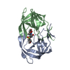
| Structure viewer | Molecule: MolmilJmol/JSmol |
|---|
- Downloads & links
-Download
| PDBx/mmCIF format | 1b2u.cif.gz | 137 KB | Display | PDBx/mmCIF format |
|---|---|---|---|---|
| PDB format | pdb1b2u.ent.gz | 107.3 KB | Display | PDB format |
| PDBx/mmJSON format | 1b2u.json.gz | Tree view | PDBx/mmJSON format | |
| Others |  Other downloads Other downloads |
-Validation report
| Arichive directory | https://data.pdbj.org/pub/pdb/validation_reports/b2/1b2uftp://data.pdbj.org/pub/pdb/validation_reports/b2/1b2u | HTTPS FTP |
|---|
-Related structure data
| Related structure data | 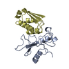 1b27C  1b2sC  1b3sC  1brsS S: Starting model for refinement C: citing same article ( |
|---|---|
| Similar structure data |


-Links
 PDBj
PDBj- Assembly
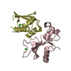
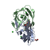
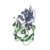
| Deposited unit | 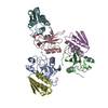
| ||||||||||||||||||||
|---|---|---|---|---|---|---|---|---|---|---|---|---|---|---|---|---|---|---|---|---|---|
| 1 | 
| ||||||||||||||||||||
| 2 | 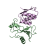
| ||||||||||||||||||||
| 3 | 
| ||||||||||||||||||||
| Unit cell |
| ||||||||||||||||||||
| Noncrystallographic symmetry (NCS) | NCS oper:
|
-Components
| #1: Protein | Mass: 12340.618 Da / Num. of mol.: 3 / Mutation: K27A Source method: isolated from a genetically manipulated source Source: (gene. exp.) #2: Protein | Mass: 10308.729 Da / Num. of mol.: 3 / Mutation: D36A Source method: isolated from a genetically manipulated source Source: (gene. exp.) #3: Water | ChemComp-HOH / |  Mass: 18.015 Da / Num. of mol.: 412 / Source method: isolated from a natural source / Formula: H2O Mass: 18.015 Da / Num. of mol.: 412 / Source method: isolated from a natural source / Formula: H2OSequence details | TER SER: THE ORIGINAL SEQUENCE OF BARSTAR OMITTED AN N-TERMINAL METHIONINE, WHICH WAS VISIBLE IN ...TER SER: THE ORIGINAL SEQUENCE OF BARSTAR OMITTED AN N-TERMINAL METHIONINE | |
|---|
-Experimental details
-Experiment
| Experiment | Method: X-RAY DIFFRACTION / Number of used crystals: 1 |
|---|
- Sample preparation
| Crystal | Density Matthews: 2.49 Å3/Da / Density % sol: 51 % Description: THE STRUCTURE WAS SOLVED BY RIGID BODY REFINEMENT OF PDB ENTRY 1BRS IN THE ASYMMETRIC UNIT | ||||||||||||||||||||
|---|---|---|---|---|---|---|---|---|---|---|---|---|---|---|---|---|---|---|---|---|---|
| Crystal grow | pH: 6.5 Details: 21% PEG-8K 0.2 M AMMONIUM SULPHATE 0.1 M NA CACODYLATE PH6.5 | ||||||||||||||||||||
| Components of the solutions |
| ||||||||||||||||||||
| Crystal grow | *PLUS Method: vapor diffusion, hanging drop | ||||||||||||||||||||
| Components of the solutions | *PLUS
|
-Data collection
| Diffraction | Mean temperature: 100 K |
|---|---|
| Diffraction source | Source: ROTATING ANODE / Type: ELLIOTT GX-13 / Wavelength: 1.5418 |
| Detector | Type: MARRESEARCH / Detector: IMAGE PLATE / Date: Jan 1, 1996 / Details: SUPER DOUBLE MIRRORS |
| Radiation | Protocol: SINGLE WAVELENGTH / Monochromatic (M) / Laue (L): M / Scattering type: x-ray |
| Radiation wavelength | Wavelength: 1.5418 Å / Relative weight: 1 |
| Reflection | Resolution: 2.1→22.3 Å / Num. obs: 32841 / % possible obs: 98.9 % / Redundancy: 3.6 % / Biso Wilson estimate: 24.56 Å2 / Rmerge(I) obs: 0.066 / Net I/σ(I): 14.45 |
| Reflection shell | Resolution: 2.1→2.15 Å / Redundancy: 3 % / Rmerge(I) obs: 0.298 / Mean I/σ(I) obs: 4 / % possible all: 98.9 |
| Reflection | *PLUS Rmerge(I) obs: 0.065 |
| Reflection shell | *PLUS % possible obs: 99.7 % |
- Processing
| Software |
| ||||||||||||||||||||||||||||||||||||||||||||||||||||||||||||||||||||||||||||||||||||
|---|---|---|---|---|---|---|---|---|---|---|---|---|---|---|---|---|---|---|---|---|---|---|---|---|---|---|---|---|---|---|---|---|---|---|---|---|---|---|---|---|---|---|---|---|---|---|---|---|---|---|---|---|---|---|---|---|---|---|---|---|---|---|---|---|---|---|---|---|---|---|---|---|---|---|---|---|---|---|---|---|---|---|---|---|---|
| Refinement | Method to determine structure: OTHER Starting model: PDB ENTRY 1BRS Resolution: 2.1→21.3 Å / SU B: 5.9 / Cross valid method: THROUGHOUT / σ(F): 0
| ||||||||||||||||||||||||||||||||||||||||||||||||||||||||||||||||||||||||||||||||||||
| Displacement parameters | Biso mean: 29.17 Å2 | ||||||||||||||||||||||||||||||||||||||||||||||||||||||||||||||||||||||||||||||||||||
| Refinement step | Cycle: LAST / Resolution: 2.1→21.3 Å
| ||||||||||||||||||||||||||||||||||||||||||||||||||||||||||||||||||||||||||||||||||||
| Refine LS restraints |
| ||||||||||||||||||||||||||||||||||||||||||||||||||||||||||||||||||||||||||||||||||||
| Software | *PLUS Name: REFMAC / Classification: refinement | ||||||||||||||||||||||||||||||||||||||||||||||||||||||||||||||||||||||||||||||||||||
| Refinement | *PLUS Highest resolution: 2.1 Å / Lowest resolution: 21.3 Å / σ(F): 0 / % reflection Rfree: 5 % / Rfactor obs: 0.214 | ||||||||||||||||||||||||||||||||||||||||||||||||||||||||||||||||||||||||||||||||||||
| Solvent computation | *PLUS | ||||||||||||||||||||||||||||||||||||||||||||||||||||||||||||||||||||||||||||||||||||
| Displacement parameters | *PLUS | ||||||||||||||||||||||||||||||||||||||||||||||||||||||||||||||||||||||||||||||||||||
| Refine LS restraints | *PLUS
|
