 Movie
Movie Controller

[English] 日本語
 Yorodumi
- PDB-2z5c: Crystal Structure of a Novel Chaperone Complex for Yeast 20S Prot... -
+ Open data

- Basic information
| Entry | Database: PDB / ID: 2z5c | ||||||
|---|---|---|---|---|---|---|---|
| Title | Crystal Structure of a Novel Chaperone Complex for Yeast 20S Proteasome Assembly | ||||||
 Components Components |
| ||||||
 Keywords Keywords | CHAPERONE/HYDROLASE / PROTEASOME / Chaperone / S. cerevisiae / CHAPERONE-HYDROLASE COMPLEX | ||||||
| Function / homology |  Function and homology information Function and homology informationnuclear-transcribed mRNA catabolic process, non-stop decay / ER-Phagosome pathway / Antigen processing: Ub, ATP-independent proteasomal degradation / Regulation of PTEN stability and activity / proteasome core complex assembly / Cross-presentation of soluble exogenous antigens (endosomes) / TNFR2 non-canonical NF-kB pathway / Proteasome assembly / CDK-mediated phosphorylation and removal of Cdc6 / FBXL7 down-regulates AURKA during mitotic entry and in early mitosis ...nuclear-transcribed mRNA catabolic process, non-stop decay / ER-Phagosome pathway / Antigen processing: Ub, ATP-independent proteasomal degradation / Regulation of PTEN stability and activity / proteasome core complex assembly / Cross-presentation of soluble exogenous antigens (endosomes) / TNFR2 non-canonical NF-kB pathway / Proteasome assembly / CDK-mediated phosphorylation and removal of Cdc6 / FBXL7 down-regulates AURKA during mitotic entry and in early mitosis / Ubiquitin-Mediated Degradation of Phosphorylated Cdc25A / KEAP1-NFE2L2 pathway / Neddylation / Orc1 removal from chromatin / MAPK6/MAPK4 signaling / Antigen processing: Ubiquitination & Proteasome degradation / Ub-specific processing proteases / proteasomal ubiquitin-independent protein catabolic process / proteasome storage granule / chaperone-mediated protein complex assembly / proteasome core complex, alpha-subunit complex / proteasome assembly / Neutrophil degranulation / proteasome complex / proteasome-mediated ubiquitin-dependent protein catabolic process / protein-containing complex / nucleus / cytoplasm Similarity search - Function | ||||||
| Biological species |  | ||||||
| Method |  X-RAY DIFFRACTION / SYNCHROTRON / MOLECULAR REPLACEMENT / Resolution: 2.9 Å X-RAY DIFFRACTION / SYNCHROTRON / MOLECULAR REPLACEMENT / Resolution: 2.9 Å | ||||||
 Authors Authors | Yashiroda, H. / Mizushima, T. / Okamoto, K. / Kameyama, T. / Hayashi, H. / Kishimoto, T. / Kasahara, M. / Kurimoto, E. / Sakata, E. / Suzuki, A. ...Yashiroda, H. / Mizushima, T. / Okamoto, K. / Kameyama, T. / Hayashi, H. / Kishimoto, T. / Kasahara, M. / Kurimoto, E. / Sakata, E. / Suzuki, A. / Hirano, Y. / Murata, S. / Kato, K. / Yamane, T. / Tanaka, K. | ||||||
 Citation Citation | Journal: Nat.Struct.Mol.Biol. / Year: 2008 Title: Crystal structure of a chaperone complex that contributes to the assembly of yeast 20S proteasomes Authors: Yashiroda, H. / Mizushima, T. / Okamoto, K. / Kameyama, T. / Hayashi, H. / Kishimoto, T. / Niwa, S. / Kasahara, M. / Kurimoto, E. / Sakata, E. / Takagi, K. / Suzuki, A. / Hirano, Y. / ...Authors: Yashiroda, H. / Mizushima, T. / Okamoto, K. / Kameyama, T. / Hayashi, H. / Kishimoto, T. / Niwa, S. / Kasahara, M. / Kurimoto, E. / Sakata, E. / Takagi, K. / Suzuki, A. / Hirano, Y. / Murata, S. / Kato, K. / Yamane, T. / Tanaka, K. | ||||||
| History |
|
- Structure visualization
| Structure viewer | Molecule: MolmilJmol/JSmol |
|---|
- Downloads & links
-Download
| PDBx/mmCIF format | 2z5c.cif.gz | 175.8 KB | Display | PDBx/mmCIF format |
|---|---|---|---|---|
| PDB format | pdb2z5c.ent.gz | 139.8 KB | Display | PDB format |
| PDBx/mmJSON format | 2z5c.json.gz | Tree view | PDBx/mmJSON format | |
| Others |  Other downloads Other downloads |
-Validation report
| Arichive directory | https://data.pdbj.org/pub/pdb/validation_reports/z5/2z5cftp://data.pdbj.org/pub/pdb/validation_reports/z5/2z5c | HTTPS FTP |
|---|
-Related structure data
| Related structure data |  2z5bSC 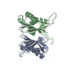 2z5eC  1rypS S: Starting model for refinement C: citing same article ( |
|---|---|
| Similar structure data |


-Links
 PDBj
PDBj


- Assembly
| Deposited unit | 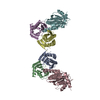
| |||||||||||||||||||||||||||||||||||||||||||||||||||||||||||||||||||||||||||||||||||||||||||||||
|---|---|---|---|---|---|---|---|---|---|---|---|---|---|---|---|---|---|---|---|---|---|---|---|---|---|---|---|---|---|---|---|---|---|---|---|---|---|---|---|---|---|---|---|---|---|---|---|---|---|---|---|---|---|---|---|---|---|---|---|---|---|---|---|---|---|---|---|---|---|---|---|---|---|---|---|---|---|---|---|---|---|---|---|---|---|---|---|---|---|---|---|---|---|---|---|---|
| 1 | 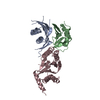
| |||||||||||||||||||||||||||||||||||||||||||||||||||||||||||||||||||||||||||||||||||||||||||||||
| 2 | 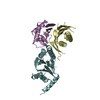
| |||||||||||||||||||||||||||||||||||||||||||||||||||||||||||||||||||||||||||||||||||||||||||||||
| Unit cell |
| |||||||||||||||||||||||||||||||||||||||||||||||||||||||||||||||||||||||||||||||||||||||||||||||
| Noncrystallographic symmetry (NCS) | NCS domain:
NCS domain segments: Component-ID: 1 / Refine code: 1
NCS ensembles :
| |||||||||||||||||||||||||||||||||||||||||||||||||||||||||||||||||||||||||||||||||||||||||||||||
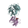
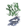
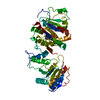
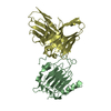
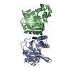
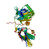
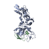
| Details | the biological unit for Dmp1 and Dmp2 is hetero dimer. In this entry, the quaternary assembly is a hetero trimer - Dmp1 and Dmp2 complexed with a part of the 20S proteasome, proteasome subunit alpha 5 |
-Components
| #1: Protein | Mass: 16876.496 Da / Num. of mol.: 2 Source method: isolated from a genetically manipulated source Source: (gene. exp.) Plasmid: pET28 / Species (production host): Escherichia coli / Production host:  #2: Protein | Mass: 16820.182 Da / Num. of mol.: 2 / Fragment: UNP residues 1-60, 91-179 Source method: isolated from a genetically manipulated source Source: (gene. exp.) Plasmid: pET28 / Species (production host): Escherichia coli / Production host: #3: Protein | Mass: 28793.217 Da / Num. of mol.: 2 Source method: isolated from a genetically manipulated source Source: (gene. exp.) Plasmid: pGEX4T / Species (production host): Escherichia coli / Production host: References: UniProt: P32379, proteasome endopeptidase complex |
|---|
-Experimental details
-Experiment
| Experiment | Method: X-RAY DIFFRACTION / Number of used crystals: 1 |
|---|
- Sample preparation
| Crystal | Density Matthews: 3.29 Å3/Da / Density % sol: 62.56 % |
|---|---|
| Crystal grow | Temperature: 293 K / Method: vapor diffusion, hanging drop / pH: 8.5 Details: 0.1M Tris-HCl, 8%(v/v) ethyleneglycol, 12%(w/v) PEG 8000, pH 8.5, VAPOR DIFFUSION, HANGING DROP, temperature 293K |
-Data collection
| Diffraction | Mean temperature: 100 K |
|---|---|
| Diffraction source | Source: SYNCHROTRON / Site: SPring-8  / Beamline: BL44XU / Wavelength: 0.9 Å / Beamline: BL44XU / Wavelength: 0.9 Å |
| Detector | Type: Bruker DIP-6040 / Detector: CCD / Date: Apr 8, 2007 |
| Radiation | Protocol: SINGLE WAVELENGTH / Monochromatic (M) / Laue (L): M / Scattering type: x-ray |
| Radiation wavelength | Wavelength: 0.9 Å / Relative weight: 1 |
| Reflection | Resolution: 2.9→50.38 Å / Num. obs: 37091 / % possible obs: 99.8 % / Observed criterion σ(I): -3 / Redundancy: 6.3 % / Rmerge(I) obs: 0.08 / Net I/σ(I): 20 |
| Reflection shell | Resolution: 2.9→3 Å / Redundancy: 6.4 % / Rmerge(I) obs: 0.35 / % possible all: 99.9 |
- Processing
| Software |
| ||||||||||||||||||||||||||||||||||||||||||||||||||||||||||||||||||||||||||||||||||||||||||||||||||||||||||||||||||||||||||||||||||||||||||||||||||||||||||||||||||||||||||
|---|---|---|---|---|---|---|---|---|---|---|---|---|---|---|---|---|---|---|---|---|---|---|---|---|---|---|---|---|---|---|---|---|---|---|---|---|---|---|---|---|---|---|---|---|---|---|---|---|---|---|---|---|---|---|---|---|---|---|---|---|---|---|---|---|---|---|---|---|---|---|---|---|---|---|---|---|---|---|---|---|---|---|---|---|---|---|---|---|---|---|---|---|---|---|---|---|---|---|---|---|---|---|---|---|---|---|---|---|---|---|---|---|---|---|---|---|---|---|---|---|---|---|---|---|---|---|---|---|---|---|---|---|---|---|---|---|---|---|---|---|---|---|---|---|---|---|---|---|---|---|---|---|---|---|---|---|---|---|---|---|---|---|---|---|---|---|---|---|---|---|---|
| Refinement | Method to determine structure: MOLECULAR REPLACEMENT Starting model: PDB ENTRY 2Z5B; PDB ENTRY 1RYP chain E Resolution: 2.9→50.38 Å / Cor.coef. Fo:Fc: 0.903 / Cor.coef. Fo:Fc free: 0.875 / SU B: 14.479 / SU ML: 0.276 / Cross valid method: THROUGHOUT / ESU R: 0.641 / ESU R Free: 0.363 / Stereochemistry target values: MAXIMUM LIKELIHOOD / Details: HYDROGENS HAVE BEEN ADDED IN THE RIDING POSITIONS
| ||||||||||||||||||||||||||||||||||||||||||||||||||||||||||||||||||||||||||||||||||||||||||||||||||||||||||||||||||||||||||||||||||||||||||||||||||||||||||||||||||||||||||
| Solvent computation | Ion probe radii: 0.8 Å / Shrinkage radii: 0.8 Å / VDW probe radii: 1.2 Å / Solvent model: MASK | ||||||||||||||||||||||||||||||||||||||||||||||||||||||||||||||||||||||||||||||||||||||||||||||||||||||||||||||||||||||||||||||||||||||||||||||||||||||||||||||||||||||||||
| Displacement parameters | Biso mean: 66.851 Å2
| ||||||||||||||||||||||||||||||||||||||||||||||||||||||||||||||||||||||||||||||||||||||||||||||||||||||||||||||||||||||||||||||||||||||||||||||||||||||||||||||||||||||||||
| Refinement step | Cycle: LAST / Resolution: 2.9→50.38 Å
| ||||||||||||||||||||||||||||||||||||||||||||||||||||||||||||||||||||||||||||||||||||||||||||||||||||||||||||||||||||||||||||||||||||||||||||||||||||||||||||||||||||||||||
| Refine LS restraints |
| ||||||||||||||||||||||||||||||||||||||||||||||||||||||||||||||||||||||||||||||||||||||||||||||||||||||||||||||||||||||||||||||||||||||||||||||||||||||||||||||||||||||||||
| Refine LS restraints NCS | Dom-ID: 1 / Refine-ID: X-RAY DIFFRACTION
| ||||||||||||||||||||||||||||||||||||||||||||||||||||||||||||||||||||||||||||||||||||||||||||||||||||||||||||||||||||||||||||||||||||||||||||||||||||||||||||||||||||||||||
| LS refinement shell | Resolution: 2.904→2.979 Å / Total num. of bins used: 20
|
