 Movie
Movie Controller


+ Open data

- Basic information
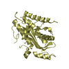
| Entry | Database: PDB / ID: 2z5e | ||||||
|---|---|---|---|---|---|---|---|
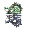
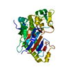
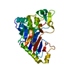
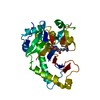
| Title | Crystal Structure of Proteasome Assembling Chaperone 3 | ||||||
 Components Components | Proteasome Assembling Chaperone 3 | ||||||
 Keywords Keywords | CHAPERONE / BETA SANDWICH / HOMODIMER / PROTEASOME | ||||||
| Function / homology |  Function and homology information Function and homology informationProteasome assembly / chaperone-mediated protein complex assembly / proteasome assembly / molecular adaptor activity / protein-containing complex binding / protein-containing complex / cytosol Similarity search - Function | ||||||
| Biological species |  Homo sapiens (human) Homo sapiens (human) | ||||||
| Method |  X-RAY DIFFRACTION / SAD / Resolution: 2 Å X-RAY DIFFRACTION / SAD / Resolution: 2 Å | ||||||
 Authors Authors | Okamoto, K. / Kurimoto, E. / Sakata, E. / Suzuki, A. / Yamane, T. / Hirano, Y. / Murata, S. / Tanaka, K. / Kato, K. | ||||||
 Citation Citation | Journal: Nat.Struct.Mol.Biol. / Year: 2008 Title: Crystal structure of a chaperone complex that contributes to the assembly of yeast 20S proteasomes Authors: Yashiroda, H. / Mizushima, T. / Okamoto, K. / Kameyama, T. / Hayashi, H. / Kishimoto, T. / Niwa, S. / Kasahara, M. / Kurimoto, E. / Sakata, E. / Takagi, K. / Suzuki, A. / Hirano, Y. / ...Authors: Yashiroda, H. / Mizushima, T. / Okamoto, K. / Kameyama, T. / Hayashi, H. / Kishimoto, T. / Niwa, S. / Kasahara, M. / Kurimoto, E. / Sakata, E. / Takagi, K. / Suzuki, A. / Hirano, Y. / Murata, S. / Kato, K. / Yamane, T. / Tanaka, K. | ||||||
| History |
|
- Structure visualization
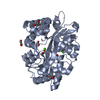
| Structure viewer | Molecule: MolmilJmol/JSmol |
|---|
- Downloads & links
-Download
| PDBx/mmCIF format | 2z5e.cif.gz | 63 KB | Display | PDBx/mmCIF format |
|---|---|---|---|---|
| PDB format | pdb2z5e.ent.gz | 47.8 KB | Display | PDB format |
| PDBx/mmJSON format | 2z5e.json.gz | Tree view | PDBx/mmJSON format | |
| Others |  Other downloads Other downloads |
-Validation report
| Arichive directory | https://data.pdbj.org/pub/pdb/validation_reports/z5/2z5eftp://data.pdbj.org/pub/pdb/validation_reports/z5/2z5e | HTTPS FTP |
|---|
-Related structure data


-Links
 PDBj
PDBj

- Assembly
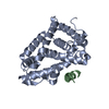
| Deposited unit | 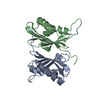
| ||||||||
|---|---|---|---|---|---|---|---|---|---|
| 1 |
| ||||||||
| Unit cell |
|
-Components
| #1: Protein | Mass: 13118.439 Da / Num. of mol.: 2 Source method: isolated from a genetically manipulated source Source: (gene. exp.) Homo sapiens (human) / Plasmid: pRSF-Duet I / Species (production host): Escherichia coli / Production host:  #2: Water | ChemComp-HOH / |  Mass: 18.015 Da / Num. of mol.: 255 / Source method: isolated from a natural source / Formula: H2O Mass: 18.015 Da / Num. of mol.: 255 / Source method: isolated from a natural source / Formula: H2O |
|---|
-Experimental details
-Experiment
| Experiment | Method: X-RAY DIFFRACTION / Number of used crystals: 1 |
|---|
- Sample preparation
| Crystal | Density Matthews: 2.14 Å3/Da / Density % sol: 42.51 % |
|---|---|
| Crystal grow | Temperature: 293.15 K / Method: vapor diffusion, hanging drop / pH: 8.5 Details: 30%(w/v) PEG4000, 0.2M MgCl2, 0.1M Tris-HCl, pH 8.5, VAPOR DIFFUSION, HANGING DROP, temperature 293.15K |
-Data collection
| Diffraction | Mean temperature: 100 K |
|---|---|
| Diffraction source | Source: ROTATING ANODE / Type: RIGAKU / Wavelength: 1.5418 Å |
| Detector | Type: RIGAKU RAXIS VII / Detector: IMAGE PLATE / Date: Jul 25, 2006 |
| Radiation | Monochromator: conforcal mirror / Protocol: SINGLE WAVELENGTH / Monochromatic (M) / Laue (L): M / Scattering type: x-ray |
| Radiation wavelength | Wavelength: 1.5418 Å / Relative weight: 1 |
| Reflection | Resolution: 2→62.99 Å / Num. all: 15971 / Num. obs: 15971 / % possible obs: 99.9 % / Observed criterion σ(I): 0 / Biso Wilson estimate: 22.7 Å2 / Rmerge(I) obs: 0.084 / Net I/σ(I): 14.9 |
| Reflection shell | Resolution: 2→2.07 Å / Redundancy: 12.4 % / Rmerge(I) obs: 0.452 / Num. unique all: 1574 / % possible all: 99.9 |
- Processing
| Software |
| ||||||||||||||||||||||||||||||||||||||||||||||||||||||||||||||||||||||||||||||||||||||||||
|---|---|---|---|---|---|---|---|---|---|---|---|---|---|---|---|---|---|---|---|---|---|---|---|---|---|---|---|---|---|---|---|---|---|---|---|---|---|---|---|---|---|---|---|---|---|---|---|---|---|---|---|---|---|---|---|---|---|---|---|---|---|---|---|---|---|---|---|---|---|---|---|---|---|---|---|---|---|---|---|---|---|---|---|---|---|---|---|---|---|---|---|
| Refinement | Method to determine structure: SAD / Resolution: 2→27.48 Å / Cor.coef. Fo:Fc: 0.955 / Cor.coef. Fo:Fc free: 0.909 / SU B: 4.407 / SU ML: 0.127 / Cross valid method: THROUGHOUT / σ(F): 0 / ESU R: 0.209 / ESU R Free: 0.193 / Stereochemistry target values: MAXIMUM LIKELIHOOD / Details: HYDROGENS HAVE BEEN ADDED IN THE RIDING POSITIONS
| ||||||||||||||||||||||||||||||||||||||||||||||||||||||||||||||||||||||||||||||||||||||||||
| Solvent computation | Ion probe radii: 0.8 Å / Shrinkage radii: 0.8 Å / VDW probe radii: 1.2 Å / Solvent model: MASK | ||||||||||||||||||||||||||||||||||||||||||||||||||||||||||||||||||||||||||||||||||||||||||
| Displacement parameters | Biso mean: 26.097 Å2
| ||||||||||||||||||||||||||||||||||||||||||||||||||||||||||||||||||||||||||||||||||||||||||
| Refine analyze |
| ||||||||||||||||||||||||||||||||||||||||||||||||||||||||||||||||||||||||||||||||||||||||||
| Refinement step | Cycle: LAST / Resolution: 2→27.48 Å
| ||||||||||||||||||||||||||||||||||||||||||||||||||||||||||||||||||||||||||||||||||||||||||
| Refine LS restraints |
| ||||||||||||||||||||||||||||||||||||||||||||||||||||||||||||||||||||||||||||||||||||||||||
| LS refinement shell | Resolution: 2→2.052 Å / Total num. of bins used: 20
|
