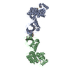
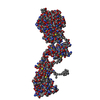
- PDB-2ylm: Mechanism of USP7 (HAUSP) activation by its C-terminal ubiquitin-... -
+
Open data
ID or keywords:
Loading...
-
Basic information
Entry
Database: PDB / ID: 2ylm
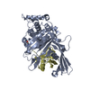
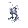
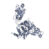
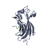
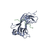
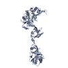
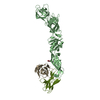
Title
Mechanism of USP7 (HAUSP) activation by its C-terminal ubiquitin-like domain (HUBL) and allosteric regulation by GMP-synthetase.
Components
UBIQUITIN CARBOXYL-TERMINAL HYDROLASE 7
Keywords
HYDROLASE / UBL
Function / homology
Function and homology information
regulation of telomere capping / regulation of establishment of protein localization to telomere / monoubiquitinated protein deubiquitination / regulation of retrograde transport, endosome to Golgi / DNA alkylation repair / deubiquitinase activity / K48-linked deubiquitinase activity / regulation of tumor necrosis factor-mediated signaling pathway / symbiont-mediated disruption of host cell PML body / negative regulation of gene expression via chromosomal CpG island methylation ...regulation of telomere capping / regulation of establishment of protein localization to telomere / monoubiquitinated protein deubiquitination / regulation of retrograde transport, endosome to Golgi / DNA alkylation repair / deubiquitinase activity / K48-linked deubiquitinase activity / regulation of tumor necrosis factor-mediated signaling pathway / symbiont-mediated disruption of host cell PML body / negative regulation of gene expression via chromosomal CpG island methylation / negative regulation of gluconeogenesis / protein deubiquitination / negative regulation of TORC1 signaling / negative regulation of proteasomal ubiquitin-dependent protein catabolic process / transcription-coupled nucleotide-excision repair / regulation of signal transduction by p53 class mediator / Regulation of PTEN localization / antiviral innate immune response / Synthesis of active ubiquitin: roles of E1 and E2 enzymes / regulation of protein stability / PML body / regulation of circadian rhythm / Transcription-Coupled Nucleotide Excision Repair (TC-NER) / Formation of TC-NER Pre-Incision Complex / p53 binding / Dual incision in TC-NER / Gap-filling DNA repair synthesis and ligation in TC-NER / Regulation of TP53 Degradation / rhythmic process / chromosome / nuclear body / ubiquitinyl hydrolase 1 / cysteine-type deubiquitinase activity / protein stabilization / Ub-specific processing proteases / protein ubiquitination / cysteine-type endopeptidase activity / protein-containing complex / proteolysis / nucleoplasm / nucleus / cytosol Similarity search - Function
In the structure databanks used in Yorodumi, some data are registered as the other names, "COVID-19 virus" and "2019-nCoV". Here are the details of the virus and the list of structure data.
Jan 31, 2019. EMDB accession codes are about to change! (news from PDBe EMDB page)
EMDB accession codes are about to change! (news from PDBe EMDB page)
The allocation of 4 digits for EMDB accession codes will soon come to an end. Whilst these codes will remain in use, new EMDB accession codes will include an additional digit and will expand incrementally as the available range of codes is exhausted. The current 4-digit format prefixed with “EMD-” (i.e. EMD-XXXX) will advance to a 5-digit format (i.e. EMD-XXXXX), and so on. It is currently estimated that the 4-digit codes will be depleted around Spring 2019, at which point the 5-digit format will come into force.
The EM Navigator/Yorodumi systems omit the EMD- prefix.
Related info.:Q: What is EMD? / ID/Accession-code notation in Yorodumi/EM Navigator
Yorodumi is a browser for structure data from EMDB, PDB, SASBDB, etc.
This page is also the successor to EM Navigator detail page, and also detail information page/front-end page for Omokage search.
The word "yorodu" (or yorozu) is an old Japanese word meaning "ten thousand". "mi" (miru) is to see.
Related info.:EMDB / PDB / SASBDB / Comparison of 3 databanks / Yorodumi Search / Aug 31, 2016. New EM Navigator & Yorodumi / Yorodumi Papers / Jmol/JSmol / Function and homology information / Changes in new EM Navigator and Yorodumi
 Movie
Movie Controller

 Yorodumi
 Open data
 Basic information
 Components
 Keywords
 Function and homology information
 HOMO SAPIENS (human)
 X-RAY DIFFRACTION /
  Authors
 Citation
 Structure visualization
 Downloads & links
 Other downloads
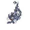


 PDBj
PDBj


 Assembly


 Mass: 35.453 Da / Num. of mol.: 1 / Source method: obtained synthetically / Formula: Cl
 Mass: 18.015 Da / Num. of mol.: 128 / Source method: isolated from a natural source / Formula: H2O
 Sample preparation
 / Beamline: ID23-2 / Wavelength: 0.873
  Processing