 Movie
Movie Controller


+ Open data

- Basic information
| Entry | Database: PDB / ID: 2f1z | ||||||
|---|---|---|---|---|---|---|---|
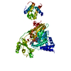
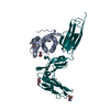
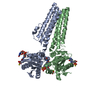
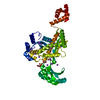
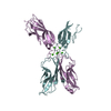
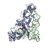
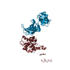
| Title | Crystal structure of HAUSP | ||||||
 Components Components | Ubiquitin carboxyl-terminal hydrolase 7 | ||||||
 Keywords Keywords | HYDROLASE / HAUSP / USP7 / UBP / deubiquitinating enzyme / substrate recognition | ||||||
| Function / homology |  Function and homology information Function and homology informationregulation of telomere capping / regulation of establishment of protein localization to telomere / monoubiquitinated protein deubiquitination / regulation of retrograde transport, endosome to Golgi / deubiquitinase activity / : / DNA alkylation repair / K48-linked deubiquitinase activity / symbiont-mediated disruption of host cell PML body / negative regulation of gene expression via chromosomal CpG island methylation ...regulation of telomere capping / regulation of establishment of protein localization to telomere / monoubiquitinated protein deubiquitination / regulation of retrograde transport, endosome to Golgi / deubiquitinase activity / : / DNA alkylation repair / K48-linked deubiquitinase activity / symbiont-mediated disruption of host cell PML body / negative regulation of gene expression via chromosomal CpG island methylation / negative regulation of gluconeogenesis / protein deubiquitination / negative regulation of TORC1 signaling / transcription-coupled nucleotide-excision repair / negative regulation of proteasomal ubiquitin-dependent protein catabolic process / Regulation of PTEN localization / regulation of signal transduction by p53 class mediator / Synthesis of active ubiquitin: roles of E1 and E2 enzymes / regulation of protein stability / regulation of circadian rhythm / PML body / Transcription-Coupled Nucleotide Excision Repair (TC-NER) / Formation of TC-NER Pre-Incision Complex / p53 binding / Dual incision in TC-NER / Gap-filling DNA repair synthesis and ligation in TC-NER / Regulation of TP53 Degradation / rhythmic process / chromosome / ubiquitinyl hydrolase 1 / cysteine-type deubiquitinase activity / protein stabilization / Ub-specific processing proteases / nuclear body / protein ubiquitination / cysteine-type endopeptidase activity / protein-containing complex / proteolysis / nucleoplasm / nucleus / cytosol Similarity search - Function | ||||||
| Biological species |  Homo sapiens (human) Homo sapiens (human) | ||||||
| Method |  X-RAY DIFFRACTION / SYNCHROTRON / MOLECULAR REPLACEMENT / Resolution: 3.2 Å X-RAY DIFFRACTION / SYNCHROTRON / MOLECULAR REPLACEMENT / Resolution: 3.2 Å | ||||||
 Authors Authors | Hu, M. / Gu, L. / Jeffrey, P.D. / Shi, Y. | ||||||
 Citation Citation | Journal: Plos Biol. / Year: 2006 Title: Structural Basis of Competitive Recognition of p53 and MDM2 by HAUSP/USP7: Implications for the Regulation of the p53-MDM2 Pathway. Authors: Hu, M. / Gu, L. / Li, M. / Jeffrey, P.D. / Gu, W. / Shi, Y. | ||||||
| History |
|
- Structure visualization
| Structure viewer | Molecule: MolmilJmol/JSmol |
|---|
- Downloads & links
-Download
| PDBx/mmCIF format | 2f1z.cif.gz | 209.8 KB | Display | PDBx/mmCIF format |
|---|---|---|---|---|
| PDB format | pdb2f1z.ent.gz | 167.7 KB | Display | PDB format |
| PDBx/mmJSON format | 2f1z.json.gz | Tree view | PDBx/mmJSON format | |
| Others |  Other downloads Other downloads |
-Validation report
| Arichive directory | https://data.pdbj.org/pub/pdb/validation_reports/f1/2f1zftp://data.pdbj.org/pub/pdb/validation_reports/f1/2f1z | HTTPS FTP |
|---|
-Related structure data
| Related structure data |  2f1wC 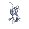 2f1xC  2f1yC 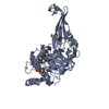 1nb8S C: citing same article ( S: Starting model for refinement |
|---|---|
| Similar structure data |


-Links
 PDBj
PDBj


- Assembly
| Deposited unit | 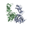
| ||||||||
|---|---|---|---|---|---|---|---|---|---|
| 1 | 
| ||||||||
| 2 | 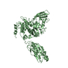
| ||||||||
| Unit cell |
|
-Components
| #1: Protein | Mass: 60532.770 Da / Num. of mol.: 2 / Fragment: residues: 43-560 Source method: isolated from a genetically manipulated source Source: (gene. exp.) Homo sapiens (human) / Gene: USP7, HAUSP / Plasmid: pGEX-2T / Species (production host): Escherichia coli / Production host:  #2: Water | ChemComp-HOH / |  Mass: 18.015 Da / Num. of mol.: 240 / Source method: isolated from a natural source / Formula: H2O Mass: 18.015 Da / Num. of mol.: 240 / Source method: isolated from a natural source / Formula: H2O |
|---|
-Experimental details
-Experiment
| Experiment | Method: X-RAY DIFFRACTION / Number of used crystals: 1 |
|---|
- Sample preparation
| Crystal | Density Matthews: 2.89 Å3/Da / Density % sol: 57.47 % |
|---|---|
| Crystal grow | Temperature: 293 K / Method: vapor diffusion, hanging drop / pH: 4.2 Details: 0.8% PEG10000, 50 mM 1,6-hexanediol, pH 4.2, VAPOR DIFFUSION, HANGING DROP, temperature 293K |
-Data collection
| Diffraction | Mean temperature: 100 K |
|---|---|
| Diffraction source | Source: SYNCHROTRON / Site: CHESS  / Beamline: A1 / Wavelength: 1.1 Å / Beamline: A1 / Wavelength: 1.1 Å |
| Detector | Type: ADSC QUANTUM 4 / Detector: CCD / Date: May 1, 2004 |
| Radiation | Monochromator: focusing mirror / Protocol: SINGLE WAVELENGTH / Monochromatic (M) / Laue (L): M / Scattering type: x-ray |
| Radiation wavelength | Wavelength: 1.1 Å / Relative weight: 1 |
| Reflection | Resolution: 3.2→99 Å / Num. all: 23732 / Num. obs: 22308 / % possible obs: 94 % / Observed criterion σ(F): 0 / Observed criterion σ(I): 0 |
| Reflection shell | Resolution: 3.2→3.3 Å / % possible all: 96.7 |
- Processing
| Software |
| ||||||||||||||||||||
|---|---|---|---|---|---|---|---|---|---|---|---|---|---|---|---|---|---|---|---|---|---|
| Refinement | Method to determine structure: MOLECULAR REPLACEMENT Starting model: PDB Entry: 1NB8 Resolution: 3.2→20 Å / σ(F): 0 / Stereochemistry target values: Engh & Huber
| ||||||||||||||||||||
| Refinement step | Cycle: LAST / Resolution: 3.2→20 Å
| ||||||||||||||||||||
| Refine LS restraints |
|
