 Movie
Movie Controller

[English] 日本語
 Yorodumi
- PDB-2f1y: Crystal structure of the TRAF-like domain of HAUSP/USP7 bound to ... -
+ Open data

- Basic information
| Entry | Database: PDB / ID: 2f1y | ||||||
|---|---|---|---|---|---|---|---|
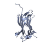
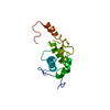
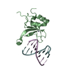
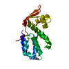
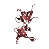
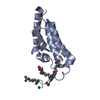
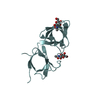
| Title | Crystal structure of the TRAF-like domain of HAUSP/USP7 bound to a MDM2 peptide | ||||||
 Components Components | HAUSP/USP7 | ||||||
 Keywords Keywords | HYDROLASE / HAUSP / USP7 / MDM2 / UBP / TRAF_like domain / MDM2 recognition / substrate binding | ||||||
| Function / homology |  Function and homology information Function and homology informationregulation of telomere capping / regulation of establishment of protein localization to telomere / cellular response to vitamin B1 / response to formaldehyde / monoubiquitinated protein deubiquitination / response to water-immersion restraint stress / response to ether / regulation of retrograde transport, endosome to Golgi / traversing start control point of mitotic cell cycle / atrial septum development ...regulation of telomere capping / regulation of establishment of protein localization to telomere / cellular response to vitamin B1 / response to formaldehyde / monoubiquitinated protein deubiquitination / response to water-immersion restraint stress / response to ether / regulation of retrograde transport, endosome to Golgi / traversing start control point of mitotic cell cycle / atrial septum development / fibroblast activation / deubiquitinase activity / regulation of protein catabolic process at postsynapse, modulating synaptic transmission / : / Trafficking of AMPA receptors / DNA alkylation repair / receptor serine/threonine kinase binding / negative regulation of intrinsic apoptotic signaling pathway by p53 class mediator / negative regulation of protein processing / positive regulation of vascular associated smooth muscle cell migration / K48-linked deubiquitinase activity / response to steroid hormone / SUMO transferase activity / peroxisome proliferator activated receptor binding / atrioventricular valve morphogenesis / AKT phosphorylates targets in the cytosol / response to iron ion / NEDD8 ligase activity / endocardial cushion morphogenesis / symbiont-mediated disruption of host cell PML body / cellular response to peptide hormone stimulus / ventricular septum development / positive regulation of muscle cell differentiation / regulation of postsynaptic neurotransmitter receptor internalization / cardiac septum morphogenesis / SUMOylation of ubiquitinylation proteins / cellular response to alkaloid / blood vessel development / negative regulation of gene expression via chromosomal CpG island methylation / Constitutive Signaling by AKT1 E17K in Cancer / ligase activity / negative regulation of DNA damage response, signal transduction by p53 class mediator / cellular response to antibiotic / SUMOylation of transcription factors / negative regulation of signal transduction by p53 class mediator / regulation of protein catabolic process / cellular response to UV-C / cellular response to estrogen stimulus / protein sumoylation / negative regulation of gluconeogenesis / response to magnesium ion / blood vessel remodeling / protein deubiquitination / ribonucleoprotein complex binding / protein localization to nucleus / protein autoubiquitination / positive regulation of vascular associated smooth muscle cell proliferation / NPAS4 regulates expression of target genes / transcription repressor complex / negative regulation of TORC1 signaling / transcription-coupled nucleotide-excision repair / positive regulation of mitotic cell cycle / negative regulation of proteasomal ubiquitin-dependent protein catabolic process / regulation of heart rate / : / positive regulation of protein export from nucleus / Regulation of PTEN localization / Synthesis of active ubiquitin: roles of E1 and E2 enzymes / regulation of signal transduction by p53 class mediator / response to cocaine / ubiquitin binding / DNA damage response, signal transduction by p53 class mediator / establishment of protein localization / sperm end piece / Stabilization of p53 / regulation of protein stability / Regulation of RUNX3 expression and activity / cellular response to gamma radiation / regulation of circadian rhythm / RING-type E3 ubiquitin transferase / PML body / Oncogene Induced Senescence / Regulation of TP53 Activity through Methylation / Degradation of CDH1 / protein destabilization / cellular response to growth factor stimulus / Transcription-Coupled Nucleotide Excision Repair (TC-NER) / response to toxic substance / Formation of TC-NER Pre-Incision Complex / cellular response to hydrogen peroxide / centriolar satellite / disordered domain specific binding / protein polyubiquitination / Dual incision in TC-NER / p53 binding / Gap-filling DNA repair synthesis and ligation in TC-NER / ubiquitin-protein transferase activity / endocytic vesicle membrane / Signaling by ALK fusions and activated point mutants / Regulation of TP53 Degradation Similarity search - Function | ||||||
| Biological species |  Homo sapiens (human) Homo sapiens (human) | ||||||
| Method |  X-RAY DIFFRACTION / SYNCHROTRON / MOLECULAR REPLACEMENT / Resolution: 1.7 Å X-RAY DIFFRACTION / SYNCHROTRON / MOLECULAR REPLACEMENT / Resolution: 1.7 Å | ||||||
 Authors Authors | Hu, M. / Gu, L. / Jeffrey, P.D. / Shi, Y. | ||||||
 Citation Citation | Journal: Plos Biol. / Year: 2006 Title: Structural Basis of Competitive Recognition of p53 and MDM2 by HAUSP/USP7: Implications for the Regulation of the p53-MDM2 Pathway. Authors: Hu, M. / Gu, L. / Li, M. / Jeffrey, P.D. / Gu, W. / Shi, Y. | ||||||
| History |
|
- Structure visualization
| Structure viewer | Molecule: MolmilJmol/JSmol |
|---|
- Downloads & links
-Download
| PDBx/mmCIF format | 2f1y.cif.gz | 45.9 KB | Display | PDBx/mmCIF format |
|---|---|---|---|---|
| PDB format | pdb2f1y.ent.gz | 31.1 KB | Display | PDB format |
| PDBx/mmJSON format | 2f1y.json.gz | Tree view | PDBx/mmJSON format | |
| Others |  Other downloads Other downloads |
-Validation report
| Arichive directory | https://data.pdbj.org/pub/pdb/validation_reports/f1/2f1yftp://data.pdbj.org/pub/pdb/validation_reports/f1/2f1y | HTTPS FTP |
|---|
-Related structure data
| Related structure data |  2f1wSC  2f1xC 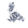 2f1zC S: Starting model for refinement C: citing same article ( |
|---|---|
| Similar structure data |


-Links
 PDBj
PDBj


- Assembly
| Deposited unit | 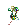
| ||||||||
|---|---|---|---|---|---|---|---|---|---|
| 1 |
| ||||||||
| Unit cell |
|
-Components
| #1: Protein | Mass: 18555.512 Da / Num. of mol.: 1 Fragment: HAUSP N-terminal domain with MDM2 peptide fused to its C-terminal Source method: isolated from a genetically manipulated source Source: (gene. exp.) Homo sapiens (human) / Plasmid: pGEX-2 / Species (production host): Escherichia coli / Production host:  |
|---|---|
| #2: Water | ChemComp-HOH /  Mass: 18.015 Da / Num. of mol.: 183 / Source method: isolated from a natural source / Formula: H2O Mass: 18.015 Da / Num. of mol.: 183 / Source method: isolated from a natural source / Formula: H2O |
-Experimental details
-Experiment
| Experiment | Method: X-RAY DIFFRACTION / Number of used crystals: 1 |
|---|
- Sample preparation
| Crystal | Density Matthews: 1.94 Å3/Da / Density % sol: 36.64 % |
|---|---|
| Crystal grow | Temperature: 293 K / Method: vapor diffusion, hanging drop / pH: 8.5 Details: 26% PEG4000, 300 mM calcium chloride, pH 8.5, VAPOR DIFFUSION, HANGING DROP, temperature 293K |
-Data collection
| Diffraction | Mean temperature: 100 K |
|---|---|
| Diffraction source | Source: SYNCHROTRON / Site: NSLS  / Beamline: X25 / Wavelength: 1.1 Å / Beamline: X25 / Wavelength: 1.1 Å |
| Detector | Type: ADSC QUANTUM 4 / Detector: CCD / Date: Feb 1, 2005 |
| Radiation | Monochromator: focusing mirror / Protocol: SINGLE WAVELENGTH / Monochromatic (M) / Laue (L): M / Scattering type: x-ray |
| Radiation wavelength | Wavelength: 1.1 Å / Relative weight: 1 |
| Reflection | Resolution: 1.7→99 Å / Num. all: 31380 / Num. obs: 30752 / % possible obs: 98 % / Observed criterion σ(F): 0 / Observed criterion σ(I): 0 |
| Reflection shell | Resolution: 1.7→1.78 Å / % possible all: 92.7 |
- Processing
| Software |
| ||||||||||||||||||||
|---|---|---|---|---|---|---|---|---|---|---|---|---|---|---|---|---|---|---|---|---|---|
| Refinement | Method to determine structure: MOLECULAR REPLACEMENT Starting model: PDB Entry: 2F1W Resolution: 1.7→20 Å / σ(F): 0 / Stereochemistry target values: Engh & Huber
| ||||||||||||||||||||
| Refinement step | Cycle: LAST / Resolution: 1.7→20 Å
| ||||||||||||||||||||
| Refine LS restraints |
|
