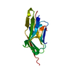
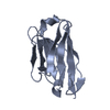
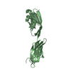
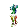
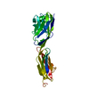
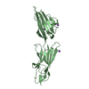
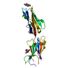
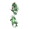
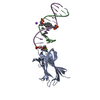
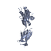
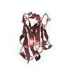
- PDB-5xj4: Complex structure of durvalumab-scFv/PD-L1 -
+
Open data
ID or keywords:
Loading...
-
Basic information
Entry
Database: PDB / ID: 5xj4
Title
Complex structure of durvalumab-scFv/PD-L1
Components
Programmed cell death 1 ligand 1
durvalumab-VH
durvalumab-VL
Keywords
IMMUNE SYSTEM / durvalumab / PD-L1 / complex structure
Function / homology
Function and homology information
negative regulation of tumor necrosis factor superfamily cytokine production / positive regulation of activated CD8-positive, alpha-beta T cell apoptotic process / negative regulation of CD8-positive, alpha-beta T cell activation / TRIF-dependent toll-like receptor signaling pathway / negative regulation of T cell mediated immune response to tumor cell / negative regulation of CD4-positive, alpha-beta T cell proliferation / STAT3 nuclear events downstream of ALK signaling / negative regulation of interleukin-10 production / negative regulation of T cell activation / negative regulation of activated T cell proliferation ...negative regulation of tumor necrosis factor superfamily cytokine production / positive regulation of activated CD8-positive, alpha-beta T cell apoptotic process / negative regulation of CD8-positive, alpha-beta T cell activation / TRIF-dependent toll-like receptor signaling pathway / negative regulation of T cell mediated immune response to tumor cell / negative regulation of CD4-positive, alpha-beta T cell proliferation / STAT3 nuclear events downstream of ALK signaling / negative regulation of interleukin-10 production / negative regulation of T cell activation / negative regulation of activated T cell proliferation / negative regulation of type II interferon production / positive regulation of interleukin-10 production / Co-inhibition by PD-1 / negative regulation of T cell receptor signaling pathway / negative regulation of T cell proliferation / T cell costimulation / response to cytokine / positive regulation of T cell proliferation / recycling endosome membrane / actin cytoskeleton / cellular response to lipopolysaccharide / early endosome membrane / adaptive immune response / transcription coactivator activity / cell surface receptor signaling pathway / immune response / receptor ligand activity / external side of plasma membrane / signal transduction / extracellular exosome / nucleoplasm / plasma membrane Similarity search - Function
Mass: 25355.795 Da / Num. of mol.: 1 Source method: isolated from a genetically manipulated source Source: (gene. exp.) Homo sapiens (human) / Gene: CD274, B7H1, PDCD1L1, PDCD1LG1, PDL1 / Production host: Escherichia coli (E. coli) / References: UniProt: Q9NZQ7
#2: Antibody
durvalumab-VH
Mass: 13422.940 Da / Num. of mol.: 1 Source method: isolated from a genetically manipulated source Source: (gene. exp.) Homo sapiens (human) / Production host: Escherichia coli (E. coli)
#3: Antibody
durvalumab-VL
Mass: 12313.643 Da / Num. of mol.: 1 Source method: isolated from a genetically manipulated source Source: (gene. exp.) Homo sapiens (human) / Production host: Escherichia coli (E. coli)
In the structure databanks used in Yorodumi, some data are registered as the other names, "COVID-19 virus" and "2019-nCoV". Here are the details of the virus and the list of structure data.
Jan 31, 2019. EMDB accession codes are about to change! (news from PDBe EMDB page)
EMDB accession codes are about to change! (news from PDBe EMDB page)
The allocation of 4 digits for EMDB accession codes will soon come to an end. Whilst these codes will remain in use, new EMDB accession codes will include an additional digit and will expand incrementally as the available range of codes is exhausted. The current 4-digit format prefixed with “EMD-” (i.e. EMD-XXXX) will advance to a 5-digit format (i.e. EMD-XXXXX), and so on. It is currently estimated that the 4-digit codes will be depleted around Spring 2019, at which point the 5-digit format will come into force.
The EM Navigator/Yorodumi systems omit the EMD- prefix.
Related info.:Q: What is EMD? / ID/Accession-code notation in Yorodumi/EM Navigator
Yorodumi is a browser for structure data from EMDB, PDB, SASBDB, etc.
This page is also the successor to EM Navigator detail page, and also detail information page/front-end page for Omokage search.
The word "yorodu" (or yorozu) is an old Japanese word meaning "ten thousand". "mi" (miru) is to see.
Related info.:EMDB / PDB / SASBDB / Comparison of 3 databanks / Yorodumi Search / Aug 31, 2016. New EM Navigator & Yorodumi / Yorodumi Papers / Jmol/JSmol / Function and homology information / Changes in new EM Navigator and Yorodumi
 Movie
Movie Controller


 Open data
 Basic information
 Components
 Keywords
 Function and homology information
 Homo sapiens (human)
 X-RAY DIFFRACTION /
  Authors
 Citation
 Structure visualization
 Downloads & links
 Other downloads
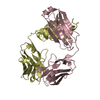
 PDBj
PDBj


 Assembly


 Mass: 18.015 Da / Num. of mol.: 169 / Source method: isolated from a natural source / Formula: H2O
 Sample preparation
 / Beamline: BL17U1 / Wavelength: 0.9791 Å
 Processing