 Movie
Movie Controller

[English] 日本語
 Yorodumi
- PDB-2zg1: Crystal Structure of Two N-terminal Domains of Siglec-5 in Comple... -
+ Open data

- Basic information
| Entry | Database: PDB / ID: 2zg1 | ||||||
|---|---|---|---|---|---|---|---|
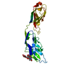
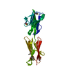
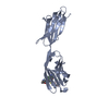
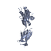
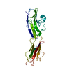
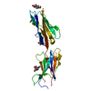
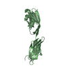
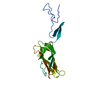
| Title | Crystal Structure of Two N-terminal Domains of Siglec-5 in Complex with 6'-Sialyllactose | ||||||
 Components Components | Sialic acid-binding Ig-like lectin 5 | ||||||
 Keywords Keywords | IMMUNE SYSTEM/CARBOHYDRATE BINDING PROTEIN / Siglec-5 inhibitory receptor / two-domain structure / V-set / C2-set / Ig-like domain / sialic acid / 6'-sialyllactose complex / Cell adhesion / Glycoprotein / Immunoglobulin domain / Lectin / Membrane / Polymorphism / Transmembrane / IMMUNE SYSTEM-CARBOHYDRATE BINDING PROTEIN COMPLEX | ||||||
| Function / homology |  Function and homology information Function and homology informationsialic acid binding / tertiary granule membrane / ficolin-1-rich granule membrane / secretory granule membrane / Immunoregulatory interactions between a Lymphoid and a non-Lymphoid cell / carbohydrate binding / cell adhesion / Neutrophil degranulation / plasma membrane Similarity search - Function | ||||||
| Biological species |  Homo sapiens (human) Homo sapiens (human) | ||||||
| Method |  X-RAY DIFFRACTION / SYNCHROTRON / MOLECULAR REPLACEMENT / Resolution: 2.7 Å X-RAY DIFFRACTION / SYNCHROTRON / MOLECULAR REPLACEMENT / Resolution: 2.7 Å | ||||||
 Authors Authors | Zhuravleva, M.A. / Sun, P.D. | ||||||
 Citation Citation | Journal: J.Mol.Biol. / Year: 2008 Title: Structural implications of Siglec-5-mediated sialoglycan recognition Authors: Zhuravleva, M.A. / Trandem, K. / Sun, P.D. | ||||||
| History |
|
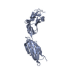
- Structure visualization
| Structure viewer | Molecule: MolmilJmol/JSmol |
|---|
- Downloads & links
-Download
| PDBx/mmCIF format | 2zg1.cif.gz | 57.2 KB | Display | PDBx/mmCIF format |
|---|---|---|---|---|
| PDB format | pdb2zg1.ent.gz | 41.5 KB | Display | PDB format |
| PDBx/mmJSON format | 2zg1.json.gz | Tree view | PDBx/mmJSON format | |
| Others |  Other downloads Other downloads |
-Validation report
| Arichive directory | https://data.pdbj.org/pub/pdb/validation_reports/zg/2zg1ftp://data.pdbj.org/pub/pdb/validation_reports/zg/2zg1 | HTTPS FTP |
|---|
-Related structure data
| Related structure data |  2zg2SC  2zg3C S: Starting model for refinement C: citing same article ( |
|---|---|
| Similar structure data |


-Links
 PDBj
PDBj


- Assembly
| Deposited unit | 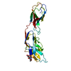
| ||||||||
|---|---|---|---|---|---|---|---|---|---|
| 1 |
| ||||||||
| Unit cell |
|
-Components
| #1: Antibody | Mass: 24332.354 Da / Num. of mol.: 1 Fragment: N-terminal V-set and C2-set domain, UNP residues 20-233 Source method: isolated from a genetically manipulated source Source: (gene. exp.) Homo sapiens (human) / Gene: SIGLEC5 / Plasmid: pET30a / Species (production host): Escherichia coli / Production host:  |
|---|---|
| #2: Sugar | ChemComp-SIA /   Type: D-saccharide, alpha linking / Mass: 309.270 Da / Num. of mol.: 1 Type: D-saccharide, alpha linking / Mass: 309.270 Da / Num. of mol.: 1Source method: isolated from a genetically manipulated source Formula: C11H19NO9 |
| #3: Water | ChemComp-HOH /  Mass: 18.015 Da / Num. of mol.: 41 / Source method: isolated from a natural source / Formula: H2O Mass: 18.015 Da / Num. of mol.: 41 / Source method: isolated from a natural source / Formula: H2O |
| Has protein modification | Y |
-Experimental details
-Experiment
| Experiment | Method: X-RAY DIFFRACTION / Number of used crystals: 1 |
|---|
- Sample preparation
| Crystal | Density Matthews: 3.67 Å3/Da / Density % sol: 66.49 % |
|---|---|
| Crystal grow | Temperature: 277 K / Method: vapor diffusion, hanging drop / pH: 8 Details: 20% MPEG 550, 0.1M TRIS, pH 8.0, VAPOR DIFFUSION, HANGING DROP, temperature 277K |
-Data collection
| Diffraction | Mean temperature: 100 K |
|---|---|
| Diffraction source | Source: SYNCHROTRON / Site: APS  / Beamline: 22-ID / Wavelength: 1 Å / Beamline: 22-ID / Wavelength: 1 Å |
| Detector | Type: MARMOSAIC 300 mm CCD / Detector: CCD / Date: Nov 12, 2005 |
| Radiation | Monochromator: GRAPHITE / Protocol: SINGLE WAVELENGTH / Monochromatic (M) / Laue (L): M / Scattering type: x-ray |
| Radiation wavelength | Wavelength: 1 Å / Relative weight: 1 |
| Reflection | Resolution: 2.7→50 Å / Num. obs: 10119 / Observed criterion σ(I): -3 / Rsym value: 0.054 / Net I/σ(I): 20.4 |
| Reflection shell | Resolution: 2.7→2.8 Å / Mean I/σ(I) obs: 3.9 / Rsym value: 0.446 |
- Processing
| Software |
| ||||||||||||||||||||||||||||||||||||||||||||||||||||||||||||||||||||||||||||||||
|---|---|---|---|---|---|---|---|---|---|---|---|---|---|---|---|---|---|---|---|---|---|---|---|---|---|---|---|---|---|---|---|---|---|---|---|---|---|---|---|---|---|---|---|---|---|---|---|---|---|---|---|---|---|---|---|---|---|---|---|---|---|---|---|---|---|---|---|---|---|---|---|---|---|---|---|---|---|---|---|---|---|
| Refinement | Method to determine structure: MOLECULAR REPLACEMENT Starting model: PDB ENTRY 2ZG2 Resolution: 2.7→25 Å / Isotropic thermal model: Isotropic / Cross valid method: THROUGHOUT / σ(F): 0 / Stereochemistry target values: Engh & Huber
| ||||||||||||||||||||||||||||||||||||||||||||||||||||||||||||||||||||||||||||||||
| Displacement parameters | Biso mean: 107 Å2 | ||||||||||||||||||||||||||||||||||||||||||||||||||||||||||||||||||||||||||||||||
| Refinement step | Cycle: LAST / Resolution: 2.7→25 Å
| ||||||||||||||||||||||||||||||||||||||||||||||||||||||||||||||||||||||||||||||||
| Refine LS restraints |
| ||||||||||||||||||||||||||||||||||||||||||||||||||||||||||||||||||||||||||||||||
| LS refinement shell | Resolution: 2.7→2.8 Å /
| ||||||||||||||||||||||||||||||||||||||||||||||||||||||||||||||||||||||||||||||||
| Xplor file |
|
