 Movie
Movie Controller

[English] 日本語
 Yorodumi
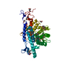
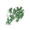
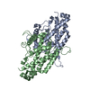
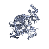
- PDB-2yfs: Crystal structure of inulosucrase from Lactobacillus johnsonii NC... -
+ Open data

- Basic information
| Entry | Database: PDB / ID: 2yfs | |||||||||
|---|---|---|---|---|---|---|---|---|---|---|
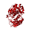
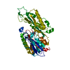
| Title | Crystal structure of inulosucrase from Lactobacillus johnsonii NCC533 in complex with sucrose | |||||||||
 Components Components | LEVANSUCRASE | |||||||||
 Keywords Keywords | TRANSFERASE / FRUCTOSYLTRANSFERASE / GLYCOSIDE HYDROLASE FAMILY GH68 / SUGAR UTILIZATION | |||||||||
| Function / homology |  Function and homology information Function and homology informationinulosucrase / inulosucrase activity / levansucrase activity / carbohydrate utilization / metal ion binding Similarity search - Function | |||||||||
| Biological species |  LACTOBACILLUS JOHNSONII (bacteria) LACTOBACILLUS JOHNSONII (bacteria) | |||||||||
| Method |  X-RAY DIFFRACTION / SYNCHROTRON / MOLECULAR REPLACEMENT / Resolution: 2.6 Å X-RAY DIFFRACTION / SYNCHROTRON / MOLECULAR REPLACEMENT / Resolution: 2.6 Å | |||||||||
 Authors Authors | Pijning, T. / Anwar, M.A. / Leemhuis, H. / Kralj, S. / Dijkhuizen, L. / Dijkstra, B.W. | |||||||||
 Citation Citation | Journal: J.Mol.Biol. / Year: 2011 Title: Crystal Structure of Inulosucrase from Lactobacillus: Insights Into the Substrate Specificity and Product Specificity of Gh68 Fructansucrases. Authors: Pijning, T. / Anwar, M.A. / Boger, M. / Dobruchowska, J.M. / Leemhuis, H. / Kralj, S. / Dijkhuizen, L. / Dijkstra, B.W. | |||||||||
| History |
|
- Structure visualization
| Structure viewer | Molecule: MolmilJmol/JSmol |
|---|
- Downloads & links
-Download
| PDBx/mmCIF format | 2yfs.cif.gz | 123.6 KB | Display | PDBx/mmCIF format |
|---|---|---|---|---|
| PDB format | pdb2yfs.ent.gz | 93.6 KB | Display | PDB format |
| PDBx/mmJSON format | 2yfs.json.gz | Tree view | PDBx/mmJSON format | |
| Others |  Other downloads Other downloads |
-Validation report
| Arichive directory | https://data.pdbj.org/pub/pdb/validation_reports/yf/2yfsftp://data.pdbj.org/pub/pdb/validation_reports/yf/2yfs | HTTPS FTP |
|---|
-Related structure data
| Related structure data | 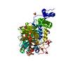 2yfrC 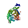 2yftC 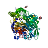 1oygS C: citing same article ( S: Starting model for refinement |
|---|---|
| Similar structure data |


-Links
 PDBj
PDBj- Assembly
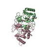
| Deposited unit | 
| ||||||||
|---|---|---|---|---|---|---|---|---|---|
| 1 |
| ||||||||
| 2 | 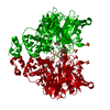
| ||||||||
| Unit cell |
|
-Components
| #1: Protein | Mass: 63852.375 Da / Num. of mol.: 1 / Fragment: RESIDUES 145-708 / Mutation: YES Source method: isolated from a genetically manipulated source Source: (gene. exp.) LACTOBACILLUS JOHNSONII (bacteria) / Strain: NCC533 / Plasmid: PETINUJ / Production host: | ||||||||
|---|---|---|---|---|---|---|---|---|---|
| #2: Polysaccharide |   Source method: isolated from a genetically manipulated source Details: oligosaccharide with reducing-end-to-reducing-end glycosidic bond References: sucrose #3: Chemical |   Mass: 40.078 Da / Num. of mol.: 2 / Source method: obtained synthetically / Formula: Ca Mass: 40.078 Da / Num. of mol.: 2 / Source method: obtained synthetically / Formula: Ca#4: Chemical | ChemComp-SO4 /   Mass: 96.063 Da / Num. of mol.: 4 / Source method: obtained synthetically / Formula: SO4 Mass: 96.063 Da / Num. of mol.: 4 / Source method: obtained synthetically / Formula: SO4#5: Water | ChemComp-HOH / |  Mass: 18.015 Da / Num. of mol.: 101 / Source method: isolated from a natural source / Formula: H2O Mass: 18.015 Da / Num. of mol.: 101 / Source method: isolated from a natural source / Formula: H2OCompound details | ENGINEERED | |
-Experimental details
-Experiment
| Experiment | Method: X-RAY DIFFRACTION / Number of used crystals: 1 |
|---|
- Sample preparation
| Crystal | Density Matthews: 3.44 Å3/Da / Density % sol: 63 % / Description: NONE |
|---|---|
| Crystal grow | pH: 6 / Details: 2M (NH4)2)SO4, 5% (V/V) 2-PROPANOL, pH 6 |
-Data collection
| Diffraction | Mean temperature: 100 K |
|---|---|
| Diffraction source | Source: SYNCHROTRON / Site: SLS  / Beamline: X06SA / Wavelength: 0.9786 / Beamline: X06SA / Wavelength: 0.9786 |
| Detector | Type: DECTRIS PILATUS 6M / Detector: PIXEL / Date: Nov 22, 2010 |
| Radiation | Protocol: SINGLE WAVELENGTH / Monochromatic (M) / Laue (L): M / Scattering type: x-ray |
| Radiation wavelength | Wavelength: 0.9786 Å / Relative weight: 1 |
| Reflection | Resolution: 2.6→39.75 Å / Num. obs: 27086 / % possible obs: 100 % / Observed criterion σ(I): -3 / Redundancy: 7.5 % / Biso Wilson estimate: 60.6 Å2 / Rmerge(I) obs: 0.1 / Net I/σ(I): 12.6 |
| Reflection shell | Resolution: 2.6→2.74 Å / Redundancy: 7.8 % / Rmerge(I) obs: 0.59 / Mean I/σ(I) obs: 3.7 / % possible all: 100 |
- Processing
| Software |
| ||||||||||||||||||||||||||||||||||||||||||||||||||||||||||||||||||||||||||||||||||||||||||||||||||||||||||||||||||||||||||||||||||||||||||||||||||||||||||||||||||||||||||||||||||||||
|---|---|---|---|---|---|---|---|---|---|---|---|---|---|---|---|---|---|---|---|---|---|---|---|---|---|---|---|---|---|---|---|---|---|---|---|---|---|---|---|---|---|---|---|---|---|---|---|---|---|---|---|---|---|---|---|---|---|---|---|---|---|---|---|---|---|---|---|---|---|---|---|---|---|---|---|---|---|---|---|---|---|---|---|---|---|---|---|---|---|---|---|---|---|---|---|---|---|---|---|---|---|---|---|---|---|---|---|---|---|---|---|---|---|---|---|---|---|---|---|---|---|---|---|---|---|---|---|---|---|---|---|---|---|---|---|---|---|---|---|---|---|---|---|---|---|---|---|---|---|---|---|---|---|---|---|---|---|---|---|---|---|---|---|---|---|---|---|---|---|---|---|---|---|---|---|---|---|---|---|---|---|---|---|
| Refinement | Method to determine structure: MOLECULAR REPLACEMENT Starting model: PDB ENTRY 1OYG Resolution: 2.6→38.45 Å / Cor.coef. Fo:Fc: 0.951 / Cor.coef. Fo:Fc free: 0.923 / SU B: 7.534 / SU ML: 0.163 / Cross valid method: THROUGHOUT / ESU R: 0.348 / ESU R Free: 0.245 / Stereochemistry target values: MAXIMUM LIKELIHOOD Details: HYDROGENS HAVE BEEN ADDED IN THE RIDING POSITIONS. RESIDUES 145-175 ARE NOT VISIBLE IN ELECTRON DENSITY
| ||||||||||||||||||||||||||||||||||||||||||||||||||||||||||||||||||||||||||||||||||||||||||||||||||||||||||||||||||||||||||||||||||||||||||||||||||||||||||||||||||||||||||||||||||||||
| Solvent computation | Ion probe radii: 0.8 Å / Shrinkage radii: 0.8 Å / VDW probe radii: 1.4 Å / Solvent model: MASK | ||||||||||||||||||||||||||||||||||||||||||||||||||||||||||||||||||||||||||||||||||||||||||||||||||||||||||||||||||||||||||||||||||||||||||||||||||||||||||||||||||||||||||||||||||||||
| Displacement parameters | Biso mean: 44.46 Å2
| ||||||||||||||||||||||||||||||||||||||||||||||||||||||||||||||||||||||||||||||||||||||||||||||||||||||||||||||||||||||||||||||||||||||||||||||||||||||||||||||||||||||||||||||||||||||
| Refinement step | Cycle: LAST / Resolution: 2.6→38.45 Å
| ||||||||||||||||||||||||||||||||||||||||||||||||||||||||||||||||||||||||||||||||||||||||||||||||||||||||||||||||||||||||||||||||||||||||||||||||||||||||||||||||||||||||||||||||||||||
| Refine LS restraints |
|