 Movie
Movie Controller

[English] 日本語
 Yorodumi
- PDB-6tn3: Crystal Structure of Aspergillus fumigatus UDP-N-acetylglucosamin... -
+ Open data

- Basic information
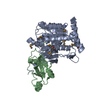
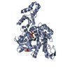
| Entry | Database: PDB / ID: 6tn3 | |||||||||
|---|---|---|---|---|---|---|---|---|---|---|
| Title | Crystal Structure of Aspergillus fumigatus UDP-N-acetylglucosamine pyrophosphorylase in complex with GlcNAc-1P | |||||||||
 Components Components | UDP-N-acetylglucosamine pyrophosphorylase | |||||||||
 Keywords Keywords | TRANSFERASE / Aspergillus fumigatus / anti-fungal / pyrophosphorylase | |||||||||
| Function / homology |  Function and homology information Function and homology informationUDP-N-acetylglucosamine diphosphorylase / UDP-N-acetylglucosamine diphosphorylase activity / UDP-N-acetylglucosamine biosynthetic process Similarity search - Function | |||||||||
| Biological species |  | |||||||||
| Method |  X-RAY DIFFRACTION / SYNCHROTRON / MOLECULAR REPLACEMENT / Resolution: 2.282 Å X-RAY DIFFRACTION / SYNCHROTRON / MOLECULAR REPLACEMENT / Resolution: 2.282 Å | |||||||||
 Authors Authors | Raimi, O.G. / Guerrero, R.H. | |||||||||
 Citation Citation | Journal: Rsc Chem Biol / Year: 2020 Title: A mechanism-inspired UDP- N -acetylglucosamine pyrophosphorylase inhibitor. Authors: Raimi, O.G. / Hurtado-Guerrero, R. / Borodkin, V. / Ferenbach, A. / Urbaniak, M.D. / Ferguson, M.A.J. / van Aalten, D.M.F. | |||||||||
| History |
|
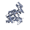
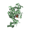
- Structure visualization
| Structure viewer | Molecule: MolmilJmol/JSmol |
|---|
- Downloads & links
-Download
| PDBx/mmCIF format | 6tn3.cif.gz | 193.8 KB | Display | PDBx/mmCIF format |
|---|---|---|---|---|
| PDB format | pdb6tn3.ent.gz | 153.7 KB | Display | PDB format |
| PDBx/mmJSON format | 6tn3.json.gz | Tree view | PDBx/mmJSON format | |
| Others |  Other downloads Other downloads |
-Validation report
| Arichive directory | https://data.pdbj.org/pub/pdb/validation_reports/tn/6tn3ftp://data.pdbj.org/pub/pdb/validation_reports/tn/6tn3 | HTTPS FTP |
|---|
-Related structure data
| Related structure data | 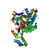 2yqsS S: Starting model for refinement |
|---|---|
| Similar structure data |


-Links
 PDBj
PDBj

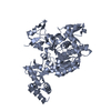
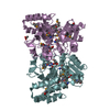
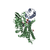
- Assembly
| Deposited unit | 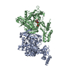
| ||||||||
|---|---|---|---|---|---|---|---|---|---|
| 1 | 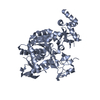
| ||||||||
| 2 | 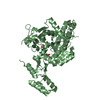
| ||||||||
| Unit cell |
|
-Components
| #1: Protein | Mass: 56793.590 Da / Num. of mol.: 2 Source method: isolated from a genetically manipulated source Source: (gene. exp.)  References: UniProt: Q4WAR0, UDP-N-acetylglucosamine diphosphorylase #2: Chemical | ChemComp-PO4 / |   Mass: 94.971 Da / Num. of mol.: 1 / Source method: obtained synthetically / Formula: PO4 Mass: 94.971 Da / Num. of mol.: 1 / Source method: obtained synthetically / Formula: PO4#3: Sugar | ChemComp-GN1 / |   Type: D-saccharide / Mass: 301.188 Da / Num. of mol.: 1 Type: D-saccharide / Mass: 301.188 Da / Num. of mol.: 1Source method: isolated from a genetically manipulated source Formula: C8H16NO9P / Feature type: SUBJECT OF INVESTIGATION #4: Water | ChemComp-HOH / |  Mass: 18.015 Da / Num. of mol.: 231 / Source method: isolated from a natural source / Formula: H2O Mass: 18.015 Da / Num. of mol.: 231 / Source method: isolated from a natural source / Formula: H2OHas ligand of interest | Y | |
|---|
-Experimental details
-Experiment
| Experiment | Method: X-RAY DIFFRACTION / Number of used crystals: 1 |
|---|
- Sample preparation
| Crystal | Density Matthews: 2.46 Å3/Da / Density % sol: 49.99 % |
|---|---|
| Crystal grow | Temperature: 277 K / Method: vapor diffusion, sitting drop / Details: 0.2 M sodium formate and 20 % PEG3350 |
-Data collection
| Diffraction | Mean temperature: 277 K / Serial crystal experiment: N |
|---|---|
| Diffraction source | Source: SYNCHROTRON / Site: ESRF  / Beamline: BM14 / Wavelength: 0.934 Å / Beamline: BM14 / Wavelength: 0.934 Å |
| Detector | Type: DECTRIS PILATUS 2M / Detector: PIXEL / Date: Aug 25, 2007 |
| Radiation | Protocol: SINGLE WAVELENGTH / Monochromatic (M) / Laue (L): M / Scattering type: x-ray |
| Radiation wavelength | Wavelength: 0.934 Å / Relative weight: 1 |
| Reflection | Resolution: 2.28→20 Å / Num. obs: 49491 / % possible obs: 95.5 % / Redundancy: 3.2 % / Biso Wilson estimate: 43.69 Å2 / Rmerge(I) obs: 0.116 / Net I/σ(I): 11.2 |
| Reflection shell | Resolution: 2.28→2.36 Å / Rmerge(I) obs: 0.392 / Num. unique obs: 4051 |
- Processing
| Software |
| ||||||||||||||||||||||||||||||||||||||||||||||||||||||||||||||||||||||||||||||||||||||||||||||||||||||||||||||||||
|---|---|---|---|---|---|---|---|---|---|---|---|---|---|---|---|---|---|---|---|---|---|---|---|---|---|---|---|---|---|---|---|---|---|---|---|---|---|---|---|---|---|---|---|---|---|---|---|---|---|---|---|---|---|---|---|---|---|---|---|---|---|---|---|---|---|---|---|---|---|---|---|---|---|---|---|---|---|---|---|---|---|---|---|---|---|---|---|---|---|---|---|---|---|---|---|---|---|---|---|---|---|---|---|---|---|---|---|---|---|---|---|---|---|---|---|
| Refinement | Method to determine structure: MOLECULAR REPLACEMENT Starting model: 2YQS Resolution: 2.282→19.925 Å / SU ML: 0.34 / Cross valid method: THROUGHOUT / σ(F): 1.34 / Phase error: 29.02
| ||||||||||||||||||||||||||||||||||||||||||||||||||||||||||||||||||||||||||||||||||||||||||||||||||||||||||||||||||
| Solvent computation | Shrinkage radii: 0.9 Å / VDW probe radii: 1.11 Å | ||||||||||||||||||||||||||||||||||||||||||||||||||||||||||||||||||||||||||||||||||||||||||||||||||||||||||||||||||
| Displacement parameters | Biso max: 102.23 Å2 / Biso mean: 48.6359 Å2 / Biso min: 20.96 Å2 | ||||||||||||||||||||||||||||||||||||||||||||||||||||||||||||||||||||||||||||||||||||||||||||||||||||||||||||||||||
| Refinement step | Cycle: final / Resolution: 2.282→19.925 Å
| ||||||||||||||||||||||||||||||||||||||||||||||||||||||||||||||||||||||||||||||||||||||||||||||||||||||||||||||||||
| Refine LS restraints |
| ||||||||||||||||||||||||||||||||||||||||||||||||||||||||||||||||||||||||||||||||||||||||||||||||||||||||||||||||||
| LS refinement shell | Refine-ID: X-RAY DIFFRACTION / Rfactor Rfree error: 0
|
