[English] 日本語
Yorodumi
- PDB-1oyg: Crystal structure of Bacillus subtilis levansucrase -

+
Open data


ID or keywords:

Loading...

-
Basic information

Entry
Database: PDB / ID: 1oyg
TitleCrystal structure of Bacillus subtilis levansucrase
Componentslevansucrase
KeywordsTRANSFERASE / glycoside hydrolase / levansucrase / beta-propeller / levan
Function / homology
Function and homology information


levansucrase / levansucrase activity / carbohydrate utilization / extracellular region / metal ion binding
Similarity search - Function
Glycoside hydrolase, family 68 / Levansucrase/Invertase / Glycosyl hydrolase domain; family 43 / 5 Propeller / Tachylectin-2; Chain A / Glycosyl hydrolase, five-bladed beta-propellor domain superfamily / Mainly Beta
Similarity search - Domain/homology
Biological speciesBacillus subtilis (bacteria)
MethodX-RAY DIFFRACTION / SYNCHROTRON / MIR / Resolution: 1.5 Å
AuthorsMeng, G. / Futterer, K.
CitationJournal: Nat.Struct.Biol. / Year: 2003
Title: Structural framework of fructosyl transfer in Bacillus subtilis levansucrase
Authors: Meng, G. / Futterer, K.
History
DepositionApr 4, 2003Deposition site: RCSB / Processing site: RCSB
Revision 1.0Oct 14, 2003Provider: repository / Type: Initial release
Revision 1.1Apr 29, 2008Group: Version format compliance
Revision 1.2Jul 13, 2011Group: Advisory / Version format compliance
Revision 1.3Feb 14, 2024Group: Data collection / Database references / Derived calculations
Category: chem_comp_atom / chem_comp_bond ...chem_comp_atom / chem_comp_bond / database_2 / pdbx_struct_conn_angle / struct_conn / struct_ref_seq_dif / struct_site
Item: _database_2.pdbx_DOI / _database_2.pdbx_database_accession ..._database_2.pdbx_DOI / _database_2.pdbx_database_accession / _pdbx_struct_conn_angle.ptnr1_auth_comp_id / _pdbx_struct_conn_angle.ptnr1_auth_seq_id / _pdbx_struct_conn_angle.ptnr1_label_asym_id / _pdbx_struct_conn_angle.ptnr1_label_atom_id / _pdbx_struct_conn_angle.ptnr1_label_comp_id / _pdbx_struct_conn_angle.ptnr1_label_seq_id / _pdbx_struct_conn_angle.ptnr3_auth_comp_id / _pdbx_struct_conn_angle.ptnr3_auth_seq_id / _pdbx_struct_conn_angle.ptnr3_label_asym_id / _pdbx_struct_conn_angle.ptnr3_label_atom_id / _pdbx_struct_conn_angle.ptnr3_label_comp_id / _pdbx_struct_conn_angle.ptnr3_label_seq_id / _pdbx_struct_conn_angle.value / _struct_conn.pdbx_dist_value / _struct_conn.ptnr1_auth_comp_id / _struct_conn.ptnr1_auth_seq_id / _struct_conn.ptnr1_label_asym_id / _struct_conn.ptnr1_label_atom_id / _struct_conn.ptnr1_label_comp_id / _struct_conn.ptnr1_label_seq_id / _struct_conn.ptnr2_auth_comp_id / _struct_conn.ptnr2_auth_seq_id / _struct_conn.ptnr2_label_asym_id / _struct_conn.ptnr2_label_atom_id / _struct_conn.ptnr2_label_comp_id / _struct_conn.ptnr2_label_seq_id / _struct_ref_seq_dif.details / _struct_site.pdbx_auth_asym_id / _struct_site.pdbx_auth_comp_id / _struct_site.pdbx_auth_seq_id

-
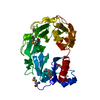
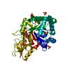
Structure visualization

Structure viewerMolecule:
MolmilJmol/JSmol

Downloads & links

-
Assembly

Deposited unit
A: levansucrase
hetero molecules


Theoretical massNumber of molelcules
Total (without water)50,7979
Polymers50,3231
Non-polymers4758
Water6,593366
1


  • Idetical with deposited unit
  • defined by author
TypeNameSymmetry operationNumber
identity operation1_555x,y,z1
Unit cell
Length a, b, c (Å)51.110, 67.014, 123.681
Angle α, β, γ (deg.)90.00, 90.00, 90.00
Int Tables number19
Space group name H-MP212121

-
Components

#1: Protein levansucrase / Beta-D-fructofuranosyl transferase / Sucrose 6-fructosyl transferase


Mass: 50322.625 Da / Num. of mol.: 1
Source method: isolated from a genetically manipulated source
Source: (gene. exp.) Bacillus subtilis (bacteria) / Gene: sacB / Plasmid: pET11c-sacBBs / Species (production host): Escherichia coli / Production host: Escherichia coli BL21(DE3) (bacteria) / Strain (production host): BL21(DE3) / References: UniProt: P05655, levansucrase
#2: Chemical ChemComp-CA / CALCIUM ION


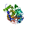
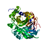
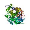
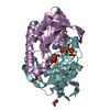
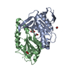
Mass: 40.078 Da / Num. of mol.: 1 / Source method: obtained synthetically / Formula: Ca
#3: Chemical
ChemComp-EDO / 1,2-ETHANEDIOL / ETHYLENE GLYCOL


Mass: 62.068 Da / Num. of mol.: 7 / Source method: obtained synthetically / Formula: C2H6O2
#4: Water ChemComp-HOH / water


Mass: 18.015 Da / Num. of mol.: 366 / Source method: isolated from a natural source / Formula: H2O

-
Experimental details

-
Experiment

ExperimentMethod: X-RAY DIFFRACTION / Number of used crystals: 1

-
Sample preparation

CrystalDensity Matthews: 2.1 Å3/Da / Density % sol: 41.54 %
Crystal grow
*PLUS
pH: 6.3 / Method: microdialysis
Components of the solutions
*PLUS
IDConc.Common nameCrystal-IDSol-IDDetails
160 mg/mlprotein11
21 mMdiammonium phosphate12
31 mMsodium acetate12pH6.3

-
Data collection

DiffractionMean temperature: 100 K
Diffraction sourceSource: SYNCHROTRON / Site: ESRF / Beamline: ID29 / Wavelength: 1.18 Å
DetectorType: ADSC QUANTUM 4 / Detector: CCD / Date: Dec 1, 2002
RadiationProtocol: SINGLE WAVELENGTH / Monochromatic (M) / Laue (L): M / Scattering type: x-ray
Radiation wavelengthWavelength: 1.18 Å / Relative weight: 1
ReflectionResolution: 1.5→30 Å / Num. obs: 67836 / % possible obs: 98.4 % / Observed criterion σ(F): 0 / Observed criterion σ(I): 0 / Redundancy: 5.1 % / Biso Wilson estimate: 9 Å2 / Rsym value: 0.091
Reflection
*PLUS
Highest resolution: 1.5 Å / Num. measured all: 683968 / Rmerge(I) obs: 0.091
Reflection shell
*PLUS
Highest resolution: 1.5 Å / Lowest resolution: 1.58 Å / % possible obs: 98.4 % / Redundancy: 3.9 % / Rmerge(I) obs: 0.095 / Mean I/σ(I) obs: 7.7

-
Processing

Software
NameVersionClassification
REFMAC5.1.19refinement
MOSFLMdata reduction
CCP4(SCALA)data scaling
SOLVEphasing
RefinementMethod to determine structure: MIR / Resolution: 1.5→30 Å / Cor.coef. Fo:Fc: 0.954 / Cor.coef. Fo:Fc free: 0.949 / SU B: 0.96 / SU ML: 0.038 / TLS residual ADP flag: LIKELY RESIDUAL / Cross valid method: THROUGHOUT / σ(F): 2 / ESU R: 0.071 / ESU R Free: 0.066 / Stereochemistry target values: MAXIMUM LIKELIHOOD
Details: THE THERMAL DISPLACEMENT PARAMETERS OF CHAIN A REFLECT THE RESIDUAL B FACTOR AND DOES NOT INCLUDE THE TLS CORRECTION.
RfactorNum. reflection% reflectionSelection details
Rfree0.17508 3386 5 %RANDOM
Rwork0.16499 ---
obs0.1655 64231 98.23 %-
all-64231 --
Solvent computationIon probe radii: 0.8 Å / Shrinkage radii: 0.8 Å / VDW probe radii: 1.4 Å / Solvent model: BABINET MODEL WITH MASK
Displacement parametersBiso mean: 4.176 Å2
Baniso -1Baniso -2Baniso -3
1--0.34 Å20 Å20 Å2
2--0.07 Å20 Å2
3---0.27 Å2
Refinement stepCycle: LAST / Resolution: 1.5→30 Å
ProteinNucleic acidLigandSolventTotal
Num. atoms3442 0 29 366 3837
Refine LS restraints
Refine-IDTypeDev idealDev ideal targetNumber
X-RAY DIFFRACTIONr_bond_refined_d0.0060.0213538
X-RAY DIFFRACTIONr_bond_other_d0.0020.022981
X-RAY DIFFRACTIONr_angle_refined_deg1.0781.9364778
X-RAY DIFFRACTIONr_angle_other_deg0.7336986
X-RAY DIFFRACTIONr_dihedral_angle_1_deg6.2855439
X-RAY DIFFRACTIONr_chiral_restr0.070.2516
X-RAY DIFFRACTIONr_gen_planes_refined0.0040.023982
X-RAY DIFFRACTIONr_gen_planes_other0.0020.02705
X-RAY DIFFRACTIONr_nbd_refined0.1970.2597
X-RAY DIFFRACTIONr_nbd_other0.2360.23520
X-RAY DIFFRACTIONr_nbtor_other0.080.21910
X-RAY DIFFRACTIONr_xyhbond_nbd_refined0.0850.2
X-RAY DIFFRACTIONr_symmetry_vdw_refined0.1350.218
X-RAY DIFFRACTIONr_symmetry_vdw_other0.2520.246
X-RAY DIFFRACTIONr_symmetry_hbond_refined0.1170.221
X-RAY DIFFRACTIONr_mcbond_it0.2911.52178
X-RAY DIFFRACTIONr_mcangle_it0.56723505
X-RAY DIFFRACTIONr_scbond_it1.05931360
X-RAY DIFFRACTIONr_scangle_it1.6564.51273
LS refinement shellResolution: 1.5→1.581 Å / Total num. of bins used: 10
RfactorNum. reflection
Rfree0.167 497
Rwork0.158 9237
obs-9734
Refinement TLS params.Method: refined / Origin x: 44.2858 Å / Origin y: 29.8237 Å / Origin z: 15.2957 Å
111213212223313233
T0.0152 Å20.0118 Å2-0.0042 Å2-0.0105 Å2-0.0091 Å2--0.0274 Å2
L0.4757 °20.0205 °20.0125 °2-0.3416 °20.049 °2--0.5204 °2
S-0.0171 Å °-0.0367 Å °0.0325 Å °0.0294 Å °0.015 Å °0.0067 Å °-0.0391 Å °0.0018 Å °0.0021 Å °
Refinement
*PLUS
Highest resolution: 1.5 Å / Lowest resolution: 30 Å / % reflection Rfree: 5 % / Rfactor Rfree: 0.175 / Rfactor Rwork: 0.165
Solvent computation
*PLUS
Displacement parameters
*PLUS
Refine LS restraints
*PLUS
Refine-IDTypeDev ideal
X-RAY DIFFRACTIONr_bond_d0.006
X-RAY DIFFRACTIONr_angle_d
X-RAY DIFFRACTIONr_angle_deg1.1
LS refinement shell
*PLUS
Highest resolution: 1.5 Å / Lowest resolution: 1.58 Å

+
About Yorodumi

-
News

-
Feb 9, 2022. New format data for meta-information of EMDB entries

New format data for meta-information of EMDB entries

  • Version 3 of the EMDB header file is now the official format.
  • The previous official version 1.9 will be removed from the archive.

Related info.:EMDB header

External links:wwPDB to switch to version 3 of the EMDB data model

-
Aug 12, 2020. Covid-19 info

Covid-19 info

URL: https://pdbjlvh1.pdbj.org/emnavi/covid19.php

New page: Covid-19 featured information page in EM Navigator.

Related info.:Covid-19 info / Mar 5, 2020. Novel coronavirus structure data

+
Mar 5, 2020. Novel coronavirus structure data

Novel coronavirus structure data

Related info.:Yorodumi Speices / Aug 12, 2020. Covid-19 info

External links:COVID-19 featured content - PDBj / Molecule of the Month (242):Coronavirus Proteases

+
Jan 31, 2019. EMDB accession codes are about to change! (news from PDBe EMDB page)

EMDB accession codes are about to change! (news from PDBe EMDB page)

  • The allocation of 4 digits for EMDB accession codes will soon come to an end. Whilst these codes will remain in use, new EMDB accession codes will include an additional digit and will expand incrementally as the available range of codes is exhausted. The current 4-digit format prefixed with “EMD-” (i.e. EMD-XXXX) will advance to a 5-digit format (i.e. EMD-XXXXX), and so on. It is currently estimated that the 4-digit codes will be depleted around Spring 2019, at which point the 5-digit format will come into force.
  • The EM Navigator/Yorodumi systems omit the EMD- prefix.

Related info.:Q: What is EMD? / ID/Accession-code notation in Yorodumi/EM Navigator

External links:EMDB Accession Codes are Changing Soon! / Contact to PDBj

+
Jul 12, 2017. Major update of PDB

Major update of PDB

  • wwPDB released updated PDB data conforming to the new PDBx/mmCIF dictionary.
  • This is a major update changing the version number from 4 to 5, and with Remediation, in which all the entries are updated.
  • In this update, many items about electron microscopy experimental information are reorganized (e.g. em_software).
  • Now, EM Navigator and Yorodumi are based on the updated data.

External links:wwPDB Remediation / Enriched Model Files Conforming to OneDep Data Standards Now Available in the PDB FTP Archive

-
Yorodumi

Thousand views of thousand structures

  • Yorodumi is a browser for structure data from EMDB, PDB, SASBDB, etc.
  • This page is also the successor to EM Navigator detail page, and also detail information page/front-end page for Omokage search.
  • The word "yorodu" (or yorozu) is an old Japanese word meaning "ten thousand". "mi" (miru) is to see.

Related info.:EMDB / PDB / SASBDB / Comparison of 3 databanks / Yorodumi Search / Aug 31, 2016. New EM Navigator & Yorodumi / Yorodumi Papers / Jmol/JSmol / Function and homology information / Changes in new EM Navigator and Yorodumi

Read more