 Movie
Movie Controller

[English] 日本語
 Yorodumi
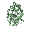
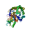
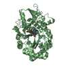
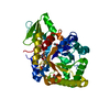
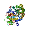
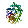
- PDB-4mnm: Crystal Structure of GH18 Chitinase (G77W/E119Q mutant) from Cyca... -
+ Open data

- Basic information
| Entry | Database: PDB / ID: 4mnm | ||||||
|---|---|---|---|---|---|---|---|
| Title | Crystal Structure of GH18 Chitinase (G77W/E119Q mutant) from Cycas revoluta in complex with (GlcNAc)4 | ||||||
 Components Components | Chitinase A | ||||||
 Keywords Keywords | HYDROLASE / Chitinase / Carbohydrate | ||||||
| Function / homology |  Function and homology information Function and homology informationchitinase activity / chitin catabolic process / chitin binding / carbohydrate metabolic process / extracellular region Similarity search - Function | ||||||
| Biological species |  | ||||||
| Method |  X-RAY DIFFRACTION / SYNCHROTRON / MOLECULAR REPLACEMENT / Resolution: 1.8 Å X-RAY DIFFRACTION / SYNCHROTRON / MOLECULAR REPLACEMENT / Resolution: 1.8 Å | ||||||
 Authors Authors | Umemoto, N. / Numata, T. / Ohnuma, T. / Osawa, T. / Taira, T. / Fukamizo, T. | ||||||
 Citation Citation | Journal: To be Published Title: Crystal Structure of GH18 Chitinase from Cycad, Cycas revoluta Authors: Umemoto, N. / Numata, T. / Ohnuma, T. / Osawa, T. / Taira, T. / Fukamizo, T. | ||||||
| History |
|
- Structure visualization
| Structure viewer | Molecule: MolmilJmol/JSmol |
|---|
- Downloads & links
-Download
| PDBx/mmCIF format | 4mnm.cif.gz | 85.7 KB | Display | PDBx/mmCIF format |
|---|---|---|---|---|
| PDB format | pdb4mnm.ent.gz | 62.9 KB | Display | PDB format |
| PDBx/mmJSON format | 4mnm.json.gz | Tree view | PDBx/mmJSON format | |
| Others |  Other downloads Other downloads |
-Validation report
| Arichive directory | https://data.pdbj.org/pub/pdb/validation_reports/mn/4mnmftp://data.pdbj.org/pub/pdb/validation_reports/mn/4mnm | HTTPS FTP |
|---|
-Related structure data
| Related structure data |  4mnjC  4mnkC  3alfS C: citing same article ( S: Starting model for refinement |
|---|---|
| Similar structure data |


-Links
 PDBj
PDBj- Assembly
| Deposited unit | 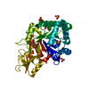
| ||||||||
|---|---|---|---|---|---|---|---|---|---|
| 1 |
| ||||||||
| Unit cell |
|
-Components
| #1: Protein | Mass: 38728.621 Da / Num. of mol.: 1 / Fragment: UNP RESIDUES 24-370 / Mutation: G77W, E119Q Source method: isolated from a genetically manipulated source Source: (gene. exp.)  | ||
|---|---|---|---|
| #2: Chemical | ChemComp-EDO /   Mass: 62.068 Da / Num. of mol.: 1 / Source method: obtained synthetically / Formula: C2H6O2 Mass: 62.068 Da / Num. of mol.: 1 / Source method: obtained synthetically / Formula: C2H6O2 | ||
| #3: Sugar | ChemComp-NAG /   Type: D-saccharide, beta linking / Mass: 221.208 Da / Num. of mol.: 1 Type: D-saccharide, beta linking / Mass: 221.208 Da / Num. of mol.: 1Source method: isolated from a genetically manipulated source Formula: C8H15NO6 | ||
| #4: Chemical | ChemComp-SO4 /   Mass: 96.063 Da / Num. of mol.: 4 / Source method: obtained synthetically / Formula: SO4 Mass: 96.063 Da / Num. of mol.: 4 / Source method: obtained synthetically / Formula: SO4#5: Water | ChemComp-HOH / |  Mass: 18.015 Da / Num. of mol.: 192 / Source method: isolated from a natural source / Formula: H2O Mass: 18.015 Da / Num. of mol.: 192 / Source method: isolated from a natural source / Formula: H2O |
-Experimental details
-Experiment
| Experiment | Method: X-RAY DIFFRACTION / Number of used crystals: 1 |
|---|
- Sample preparation
| Crystal | Density Matthews: 2.1 Å3/Da / Density % sol: 41.34 % |
|---|---|
| Crystal grow | Temperature: 293 K / Method: vapor diffusion, sitting drop / pH: 6.5 Details: 0.01M Magnesium sulfate heptahydrate, 0.05M Sodium cacodylate trihydrate pH 6.5, 2.0M Ammonium sulfate, VAPOR DIFFUSION, SITTING DROP, temperature 293K |
-Data collection
| Diffraction | Mean temperature: 95 K |
|---|---|
| Diffraction source | Source: SYNCHROTRON / Site: Photon Factory  / Beamline: BL-17A / Wavelength: 0.98 Å / Beamline: BL-17A / Wavelength: 0.98 Å |
| Detector | Type: ADSC QUANTUM 270 / Detector: CCD / Date: Feb 23, 2013 |
| Radiation | Monochromator: Numerical link type Si(111) double crystal monochromator, liquid nitrogen cooling Protocol: SINGLE WAVELENGTH / Monochromatic (M) / Laue (L): M / Scattering type: x-ray |
| Radiation wavelength | Wavelength: 0.98 Å / Relative weight: 1 |
| Reflection | Resolution: 1.8→51.61 Å / Num. obs: 30778 / % possible obs: 99.7 % / Redundancy: 7.2 % / Biso Wilson estimate: 15 Å2 / Rmerge(I) obs: 0.07 / Net I/σ(I): 43.3 |
| Reflection shell | Resolution: 1.8→1.83 Å / Redundancy: 7.2 % / Rmerge(I) obs: 0.335 / Mean I/σ(I) obs: 10.6 / Num. unique all: 10829 / % possible all: 99.7 |
- Processing
| Software |
| |||||||||||||||||||||||||||||||||||||||||||||||||||||||||||||||||
|---|---|---|---|---|---|---|---|---|---|---|---|---|---|---|---|---|---|---|---|---|---|---|---|---|---|---|---|---|---|---|---|---|---|---|---|---|---|---|---|---|---|---|---|---|---|---|---|---|---|---|---|---|---|---|---|---|---|---|---|---|---|---|---|---|---|---|
| Refinement | Method to determine structure: MOLECULAR REPLACEMENT Starting model: PDB ENTRY 3ALF Resolution: 1.8→50 Å / Cor.coef. Fo:Fc: 0.943 / Cor.coef. Fo:Fc free: 0.93 / SU B: 2.532 / SU ML: 0.081 / Isotropic thermal model: RESTRAINED / Cross valid method: THROUGHOUT / ESU R: 0.145 / ESU R Free: 0.127 / Stereochemistry target values: MAXIMUM LIKELIHOOD
| |||||||||||||||||||||||||||||||||||||||||||||||||||||||||||||||||
| Solvent computation | Ion probe radii: 0.8 Å / Shrinkage radii: 0.8 Å / VDW probe radii: 1.4 Å / Solvent model: MASK | |||||||||||||||||||||||||||||||||||||||||||||||||||||||||||||||||
| Displacement parameters | Biso mean: 14.451 Å2
| |||||||||||||||||||||||||||||||||||||||||||||||||||||||||||||||||
| Refinement step | Cycle: LAST / Resolution: 1.8→50 Å
| |||||||||||||||||||||||||||||||||||||||||||||||||||||||||||||||||
| Refine LS restraints |
| |||||||||||||||||||||||||||||||||||||||||||||||||||||||||||||||||
| LS refinement shell | Resolution: 1.801→1.848 Å / Total num. of bins used: 20
|
