 Movie
Movie Controller

[English] 日本語
 Yorodumi
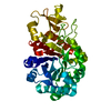
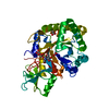
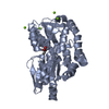
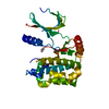
- PDB-3aqu: Crystal structure of a class V chitinase from Arabidopsis thaliana -
+ Open data

- Basic information
| Entry | Database: PDB / ID: 3aqu | ||||||
|---|---|---|---|---|---|---|---|
| Title | Crystal structure of a class V chitinase from Arabidopsis thaliana | ||||||
 Components Components | At4g19810 | ||||||
 Keywords Keywords | HYDROLASE / stress response / TIM barrel / chitin | ||||||
| Function / homology |  Function and homology information Function and homology informationexo-chitinase (non-reducing end) / exochitinase activity / secretory vesicle / chitinase activity / plant-type cell wall / response to jasmonic acid / response to abscisic acid / endochitinase activity / chitinase / chitin catabolic process ...exo-chitinase (non-reducing end) / exochitinase activity / secretory vesicle / chitinase activity / plant-type cell wall / response to jasmonic acid / response to abscisic acid / endochitinase activity / chitinase / chitin catabolic process / chitin binding / polysaccharide catabolic process / response to salt stress / extracellular region Similarity search - Function | ||||||
| Biological species |  | ||||||
| Method |  X-RAY DIFFRACTION / SYNCHROTRON / MOLECULAR REPLACEMENT / Resolution: 2.01 Å X-RAY DIFFRACTION / SYNCHROTRON / MOLECULAR REPLACEMENT / Resolution: 2.01 Å | ||||||
 Authors Authors | Numata, T. / Ohnuma, T. / Osawa, T. / Fukamizo, T. | ||||||
 Citation Citation | Journal: Planta / Year: 2011 Title: A class V chitinase from Arabidopsis thaliana: gene responses, enzymatic properties, and crystallographic analysis Authors: Ohnuma, T. / Numata, T. / Osawa, T. / Mizuhara, M. / Lampela, O. / Juffer, A.H. / Skriver, K. / Fukamizo, T. | ||||||
| History |
|
- Structure visualization
| Structure viewer | Molecule: MolmilJmol/JSmol |
|---|
- Downloads & links
-Download
| PDBx/mmCIF format | 3aqu.cif.gz | 275.5 KB | Display | PDBx/mmCIF format |
|---|---|---|---|---|
| PDB format | pdb3aqu.ent.gz | 223 KB | Display | PDB format |
| PDBx/mmJSON format | 3aqu.json.gz | Tree view | PDBx/mmJSON format | |
| Others |  Other downloads Other downloads |
-Validation report
| Arichive directory | https://data.pdbj.org/pub/pdb/validation_reports/aq/3aquftp://data.pdbj.org/pub/pdb/validation_reports/aq/3aqu | HTTPS FTP |
|---|
-Related structure data
| Related structure data |  3alfS S: Starting model for refinement |
|---|---|
| Similar structure data |


-Links
 PDBj
PDBj


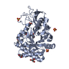
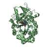
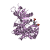
- Assembly
| Deposited unit | 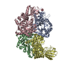
| ||||||||
|---|---|---|---|---|---|---|---|---|---|
| 1 | 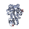
| ||||||||
| 2 | 
| ||||||||
| 3 | 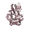
| ||||||||
| 4 | 
| ||||||||
| Unit cell |
|
-Components
| #1: Protein | Mass: 38673.824 Da / Num. of mol.: 4 / Fragment: residues in UNP 25-379 Source method: isolated from a genetically manipulated source Source: (gene. exp.)  #2: Chemical |   Mass: 189.100 Da / Num. of mol.: 3 / Source method: obtained synthetically / Formula: C6H5O7 Mass: 189.100 Da / Num. of mol.: 3 / Source method: obtained synthetically / Formula: C6H5O7#3: Water | ChemComp-HOH / |  Mass: 18.015 Da / Num. of mol.: 581 / Source method: isolated from a natural source / Formula: H2O Mass: 18.015 Da / Num. of mol.: 581 / Source method: isolated from a natural source / Formula: H2O |
|---|
-Experimental details
-Experiment
| Experiment | Method: X-RAY DIFFRACTION / Number of used crystals: 1 |
|---|
- Sample preparation
| Crystal | Density Matthews: 3.08 Å3/Da / Density % sol: 60.03 % |
|---|---|
| Crystal grow | Temperature: 293 K / Method: vapor diffusion, sitting drop / pH: 5.6 Details: 100mM trisodium citrate, 20% PEG10000, pH 5.6, VAPOR DIFFUSION, SITTING DROP, temperature 293K |
-Data collection
| Diffraction | Mean temperature: 95 K |
|---|---|
| Diffraction source | Source: SYNCHROTRON / Site: Photon Factory  / Beamline: BL-17A / Wavelength: 0.98 Å / Beamline: BL-17A / Wavelength: 0.98 Å |
| Detector | Type: ADSC QUANTUM 270 / Detector: CCD / Date: Mar 17, 2010 / Details: mirrors |
| Radiation | Monochromator: Numerical link type Si(111) double crystal monochromator, liquid nitrogen cooling Protocol: SINGLE WAVELENGTH / Monochromatic (M) / Laue (L): M / Scattering type: x-ray |
| Radiation wavelength | Wavelength: 0.98 Å / Relative weight: 1 |
| Reflection | Resolution: 2→50 Å / Num. obs: 116512 / % possible obs: 94.5 % / Redundancy: 2.9 % / Rsym value: 0.102 / Net I/σ(I): 7.6 |
| Reflection shell | Resolution: 2→2.03 Å / Redundancy: 2.3 % / Rmerge(I) obs: 0.336 / Mean I/σ(I) obs: 1.9 / % possible all: 88.7 |
- Processing
| Software |
| |||||||||||||||||||||||||||||||||||||||||||||||||||||||||||||||||
|---|---|---|---|---|---|---|---|---|---|---|---|---|---|---|---|---|---|---|---|---|---|---|---|---|---|---|---|---|---|---|---|---|---|---|---|---|---|---|---|---|---|---|---|---|---|---|---|---|---|---|---|---|---|---|---|---|---|---|---|---|---|---|---|---|---|---|
| Refinement | Method to determine structure: MOLECULAR REPLACEMENT Starting model: 3ALF Resolution: 2.01→50 Å / Cor.coef. Fo:Fc: 0.923 / Cor.coef. Fo:Fc free: 0.898 / SU B: 3.869 / SU ML: 0.106 / Cross valid method: THROUGHOUT / ESU R Free: 0.15 / Stereochemistry target values: MAXIMUM LIKELIHOOD / Details: HYDROGENS HAVE BEEN ADDED IN THE RIDING POSITIONS
| |||||||||||||||||||||||||||||||||||||||||||||||||||||||||||||||||
| Solvent computation | Ion probe radii: 0.8 Å / Shrinkage radii: 0.8 Å / VDW probe radii: 1.4 Å / Solvent model: MASK | |||||||||||||||||||||||||||||||||||||||||||||||||||||||||||||||||
| Displacement parameters | Biso mean: 12.842 Å2
| |||||||||||||||||||||||||||||||||||||||||||||||||||||||||||||||||
| Refinement step | Cycle: LAST / Resolution: 2.01→50 Å
| |||||||||||||||||||||||||||||||||||||||||||||||||||||||||||||||||
| Refine LS restraints |
| |||||||||||||||||||||||||||||||||||||||||||||||||||||||||||||||||
| LS refinement shell | Resolution: 2.005→2.057 Å / Total num. of bins used: 20
|
