 Movie
Movie Controller

[English] 日本語
 Yorodumi
- PDB-4g1p: Structural and Mechanistic Basis of Substrate Recognition by Nove... -
+ Open data

- Basic information
| Entry | Database: PDB / ID: 4g1p | ||||||
|---|---|---|---|---|---|---|---|
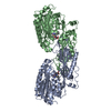
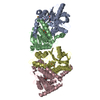
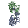
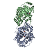
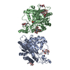
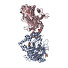
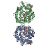
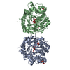
| Title | Structural and Mechanistic Basis of Substrate Recognition by Novel Di-peptidase Dug1p From Saccharomyces cerevisiae | ||||||
 Components Components | Cys-Gly metallodipeptidase DUG1 | ||||||
 Keywords Keywords | HYDROLASE / Di-nuclear peptidases / M20 family metallo-peptidases / alpha/beta scaffold / N-terminal catalytic domain/C-terminal lid domain | ||||||
| Function / homology |  Function and homology information Function and homology informationGlutathione synthesis and recycling / Paracetamol ADME / Hydrolases; Acting on peptide bonds (peptidases); Dipeptidases / glutathione catabolic process / metallodipeptidase activity / peptidase activity / omega peptidase activity / mitochondrion / proteolysis / metal ion binding ...Glutathione synthesis and recycling / Paracetamol ADME / Hydrolases; Acting on peptide bonds (peptidases); Dipeptidases / glutathione catabolic process / metallodipeptidase activity / peptidase activity / omega peptidase activity / mitochondrion / proteolysis / metal ion binding / identical protein binding / cytoplasm Similarity search - Function | ||||||
| Biological species |  | ||||||
| Method |  X-RAY DIFFRACTION / MOLECULAR REPLACEMENT / Resolution: 2.547 Å X-RAY DIFFRACTION / MOLECULAR REPLACEMENT / Resolution: 2.547 Å | ||||||
 Authors Authors | Singh, A.K. / Singh, M. / Pandya, V.K. / Singh, V. / Mittal, M. / Kumaran, S. | ||||||
 Citation Citation | Journal: To be published Title: Structural and Mechanistic Basis of Substrate Recognition by Novel Di-peptidase Dug1p From Saccromyces cerevesiae Authors: Singh, A.K. / Singh, M. / Pandya, V.K. / Singh, V. / Mittal, M. / Kumaran, S. | ||||||
| History |
|
- Structure visualization
| Structure viewer | Molecule: MolmilJmol/JSmol |
|---|
- Downloads & links
-Download
| PDBx/mmCIF format | 4g1p.cif.gz | 109.7 KB | Display | PDBx/mmCIF format |
|---|---|---|---|---|
| PDB format | pdb4g1p.ent.gz | 83 KB | Display | PDB format |
| PDBx/mmJSON format | 4g1p.json.gz | Tree view | PDBx/mmJSON format | |
| Others |  Other downloads Other downloads |
-Validation report
| Arichive directory | https://data.pdbj.org/pub/pdb/validation_reports/g1/4g1pftp://data.pdbj.org/pub/pdb/validation_reports/g1/4g1p | HTTPS FTP |
|---|
-Related structure data
| Related structure data |  2zofS S: Starting model for refinement |
|---|---|
| Similar structure data |
-Links
 PDBj
PDBj


- Assembly
| Deposited unit | 
| ||||||||
|---|---|---|---|---|---|---|---|---|---|
| 1 | 
| ||||||||
| Unit cell |
|
-Components
| #1: Protein | Mass: 53760.027 Da / Num. of mol.: 1 Source method: isolated from a genetically manipulated source Source: (gene. exp.) Strain: S288c / Gene: Dug1p / Plasmid: pET23a / Production host:  References: UniProt: P43616, Hydrolases; Acting on peptide bonds (peptidases); Dipeptidases | ||||||||
|---|---|---|---|---|---|---|---|---|---|
| #2: Chemical |   Mass: 65.409 Da / Num. of mol.: 2 / Source method: obtained synthetically / Formula: Zn Mass: 65.409 Da / Num. of mol.: 2 / Source method: obtained synthetically / Formula: Zn#3: Chemical | ChemComp-GLY / |   Type: peptide linking / Mass: 75.067 Da / Num. of mol.: 1 / Source method: obtained synthetically / Formula: C2H5NO2 Type: peptide linking / Mass: 75.067 Da / Num. of mol.: 1 / Source method: obtained synthetically / Formula: C2H5NO2#4: Chemical | ChemComp-CYS / |   Type: L-peptide linking / Mass: 121.158 Da / Num. of mol.: 1 / Source method: obtained synthetically / Formula: C3H7NO2S Type: L-peptide linking / Mass: 121.158 Da / Num. of mol.: 1 / Source method: obtained synthetically / Formula: C3H7NO2S#5: Water | ChemComp-HOH / |  Mass: 18.015 Da / Num. of mol.: 168 / Source method: isolated from a natural source / Formula: H2O Mass: 18.015 Da / Num. of mol.: 168 / Source method: isolated from a natural source / Formula: H2ONonpolymer details | DI-PEPTIDE, GLY-CYS IS INTRINSIC DIPEPTIDE. | |
-Experimental details
-Experiment
| Experiment | Method: X-RAY DIFFRACTION / Number of used crystals: 1 |
|---|
- Sample preparation
| Crystal | Density Matthews: 3.41 Å3/Da / Density % sol: 63.95 % |
|---|---|
| Crystal grow | Temperature: 291 K / Method: vapor diffusion, sitting drop / pH: 5.6 Details: 0.5 M AmSO4, 0.1 M tri-Na citrate pH 5.6, 1.0 M Lithium sulfate, VAPOR DIFFUSION, SITTING DROP, temperature 291K |
-Data collection
| Diffraction | Mean temperature: 100 K |
|---|---|
| Diffraction source | Source: ROTATING ANODE / Type: RIGAKU MICROMAX-007 HF / Wavelength: 1.542 Å |
| Detector | Type: MAR scanner 345 mm plate / Detector: IMAGE PLATE / Date: Aug 16, 2010 |
| Radiation | Monochromator: mirror / Protocol: SINGLE WAVELENGTH / Monochromatic (M) / Laue (L): M / Scattering type: x-ray |
| Radiation wavelength | Wavelength: 1.542 Å / Relative weight: 1 |
| Reflection | Resolution: 2.547→50 Å / Num. obs: 24507 / Observed criterion σ(I): 2 / Redundancy: 11.3 % / Biso Wilson estimate: 36.42 Å2 / Rmerge(I) obs: 0.216 |
| Reflection shell | Resolution: 2.55→2.7 Å / Redundancy: 11.21 % / Rmerge(I) obs: 0.874 / Mean I/σ(I) obs: 2.27 |
- Processing
| Software |
| ||||||||||||||||||||||||||||||||||||||||||||||||||||||||||||||||||||||
|---|---|---|---|---|---|---|---|---|---|---|---|---|---|---|---|---|---|---|---|---|---|---|---|---|---|---|---|---|---|---|---|---|---|---|---|---|---|---|---|---|---|---|---|---|---|---|---|---|---|---|---|---|---|---|---|---|---|---|---|---|---|---|---|---|---|---|---|---|---|---|---|
| Refinement | Method to determine structure: MOLECULAR REPLACEMENT Starting model: 2ZOF Resolution: 2.547→19.731 Å / Occupancy max: 1 / Occupancy min: 1 / FOM work R set: 0.841 / SU ML: 0.32 / σ(F): 1.99 / Phase error: 22.11 / Stereochemistry target values: ML
| ||||||||||||||||||||||||||||||||||||||||||||||||||||||||||||||||||||||
| Solvent computation | Shrinkage radii: 0.9 Å / VDW probe radii: 1.11 Å / Solvent model: FLAT BULK SOLVENT MODEL | ||||||||||||||||||||||||||||||||||||||||||||||||||||||||||||||||||||||
| Displacement parameters | Biso max: 83.74 Å2 / Biso mean: 36.42 Å2 / Biso min: 10.82 Å2 | ||||||||||||||||||||||||||||||||||||||||||||||||||||||||||||||||||||||
| Refinement step | Cycle: LAST / Resolution: 2.547→19.731 Å
| ||||||||||||||||||||||||||||||||||||||||||||||||||||||||||||||||||||||
| Refine LS restraints |
| ||||||||||||||||||||||||||||||||||||||||||||||||||||||||||||||||||||||
| LS refinement shell | Refine-ID: X-RAY DIFFRACTION / Total num. of bins used: 9
|
