 Movie
Movie Controller


+ Open data

- Basic information
| Entry | Database: PDB / ID: 1zi7 | ||||||
|---|---|---|---|---|---|---|---|
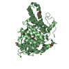
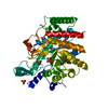
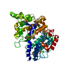
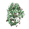
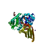
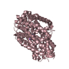
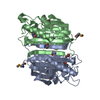
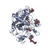
| Title | Structure of truncated yeast oxysterol binding protein Osh4 | ||||||
 Components Components | KES1 protein | ||||||
 Keywords Keywords | LIPID BINDING PROTEIN / oxysterol / sterol binding protein | ||||||
| Function / homology |  Function and homology information Function and homology informationAcyl chain remodelling of PS / Synthesis of bile acids and bile salts / ER to Golgi ceramide transport / sterol transfer activity / post-Golgi vesicle-mediated transport / sterol transport / sphingolipid metabolic process / maintenance of cell polarity / piecemeal microautophagy of the nucleus / oxysterol binding ...Acyl chain remodelling of PS / Synthesis of bile acids and bile salts / ER to Golgi ceramide transport / sterol transfer activity / post-Golgi vesicle-mediated transport / sterol transport / sphingolipid metabolic process / maintenance of cell polarity / piecemeal microautophagy of the nucleus / oxysterol binding / phosphatidic acid binding / phosphatidylinositol-4-phosphate binding / exocytosis / phosphatidylinositol-4,5-bisphosphate binding / endocytosis / Golgi membrane / lipid binding / membrane / cytoplasm / cytosol Similarity search - Function | ||||||
| Biological species |  | ||||||
| Method |  X-RAY DIFFRACTION / SYNCHROTRON / MOLECULAR REPLACEMENT / Resolution: 2.5 Å X-RAY DIFFRACTION / SYNCHROTRON / MOLECULAR REPLACEMENT / Resolution: 2.5 Å | ||||||
 Authors Authors | Im, Y.J. / Raychaudhuri, S. / Prinz, W.A. / Hurley, J.H. | ||||||
 Citation Citation | Journal: Nature / Year: 2005 Title: Structural mechanism for sterol sensing and transport by OSBP-related proteins Authors: Im, Y.J. / Raychaudhuri, S. / Prinz, W.A. / Hurley, J.H. | ||||||
| History |
|
- Structure visualization
| Structure viewer | Molecule: MolmilJmol/JSmol |
|---|
- Downloads & links
-Download
| PDBx/mmCIF format | 1zi7.cif.gz | 248.9 KB | Display | PDBx/mmCIF format |
|---|---|---|---|---|
| PDB format | pdb1zi7.ent.gz | 201.3 KB | Display | PDB format |
| PDBx/mmJSON format | 1zi7.json.gz | Tree view | PDBx/mmJSON format | |
| Others |  Other downloads Other downloads |
-Validation report
| Arichive directory | https://data.pdbj.org/pub/pdb/validation_reports/zi/1zi7ftp://data.pdbj.org/pub/pdb/validation_reports/zi/1zi7 | HTTPS FTP |
|---|
-Related structure data
| Related structure data | 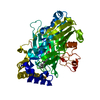 1zhtC  1zhwC 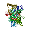 1zhxSC 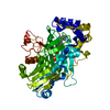 1zhyC  1zhzC C: citing same article ( S: Starting model for refinement |
|---|---|
| Similar structure data |


-Links
 PDBj
PDBj- Assembly
| Deposited unit | 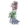
| ||||||||
|---|---|---|---|---|---|---|---|---|---|
| 1 | 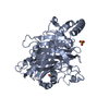
| ||||||||
| 2 | 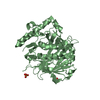
| ||||||||
| 3 | 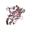
| ||||||||
| 4 | 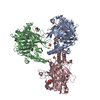
| ||||||||
| 5 | 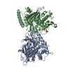
| ||||||||
| 6 | 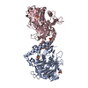
| ||||||||
| Unit cell |
|
-Components
| #1: Protein | Mass: 46491.566 Da / Num. of mol.: 3 / Fragment: Residues 30 - 434 / Mutation: 236RGYFS -> VD Source method: isolated from a genetically manipulated source Source: (gene. exp.) Gene: OSH4 / Plasmid: modified pGEX4T / Species (production host): Escherichia coli / Production host:  #2: Chemical | ChemComp-SO4 /   Mass: 96.063 Da / Num. of mol.: 6 / Source method: obtained synthetically / Formula: SO4 Mass: 96.063 Da / Num. of mol.: 6 / Source method: obtained synthetically / Formula: SO4#3: Water | ChemComp-HOH / |  Mass: 18.015 Da / Num. of mol.: 141 / Source method: isolated from a natural source / Formula: H2O Mass: 18.015 Da / Num. of mol.: 141 / Source method: isolated from a natural source / Formula: H2O |
|---|
-Experimental details
-Experiment
| Experiment | Method: X-RAY DIFFRACTION / Number of used crystals: 1 |
|---|
- Sample preparation
| Crystal | Density Matthews: 4 Å3/Da / Density % sol: 69 % |
|---|---|
| Crystal grow | Temperature: 298 K / Method: vapor diffusion, hanging drop / pH: 6 Details: Ammonium sulfate, MES, pH 6.0, VAPOR DIFFUSION, HANGING DROP, temperature 298.0K |
-Data collection
| Diffraction | Mean temperature: 298 K |
|---|---|
| Diffraction source | Source: SYNCHROTRON / Site: APS  / Beamline: 22-ID / Wavelength: 0.9792 / Wavelength: 0.9792 Å / Beamline: 22-ID / Wavelength: 0.9792 / Wavelength: 0.9792 Å |
| Detector | Type: MARRESEARCH / Detector: CCD / Date: Feb 6, 2004 |
| Radiation | Protocol: SINGLE WAVELENGTH / Monochromatic (M) / Laue (L): M / Scattering type: x-ray |
| Radiation wavelength | Wavelength: 0.9792 Å / Relative weight: 1 |
| Reflection | Resolution: 2.5→50 Å / Num. all: 78298 / Num. obs: 75232 / % possible obs: 99.3 % / Observed criterion σ(F): 0 / Observed criterion σ(I): 3 / Redundancy: 3.3 % / Biso Wilson estimate: 39.2 Å2 / Rsym value: 0.066 / Net I/σ(I): 22.9 |
| Reflection shell | Resolution: 2.5→2.59 Å / Mean I/σ(I) obs: 3.16 / Num. unique all: 8145 / Rsym value: 0.355 / % possible all: 96.2 |
- Processing
| Software |
| ||||||||||||||||||||||||||||||||||||||||||||||||||||||||||||
|---|---|---|---|---|---|---|---|---|---|---|---|---|---|---|---|---|---|---|---|---|---|---|---|---|---|---|---|---|---|---|---|---|---|---|---|---|---|---|---|---|---|---|---|---|---|---|---|---|---|---|---|---|---|---|---|---|---|---|---|---|---|
| Refinement | Method to determine structure: MOLECULAR REPLACEMENT Starting model: PDB entry 1ZHX Resolution: 2.5→45.11 Å / Rfactor Rfree error: 0.004 / Data cutoff high absF: 1816335.14 / Data cutoff low absF: 0 / Isotropic thermal model: RESTRAINED / Cross valid method: THROUGHOUT / σ(F): 0 / Stereochemistry target values: Engh & Huber
| ||||||||||||||||||||||||||||||||||||||||||||||||||||||||||||
| Solvent computation | Solvent model: FLAT MODEL / Bsol: 33.908 Å2 / ksol: 0.370563 e/Å3 | ||||||||||||||||||||||||||||||||||||||||||||||||||||||||||||
| Displacement parameters | Biso mean: 48.4 Å2
| ||||||||||||||||||||||||||||||||||||||||||||||||||||||||||||
| Refine analyze |
| ||||||||||||||||||||||||||||||||||||||||||||||||||||||||||||
| Refinement step | Cycle: LAST / Resolution: 2.5→45.11 Å
| ||||||||||||||||||||||||||||||||||||||||||||||||||||||||||||
| Refine LS restraints |
| ||||||||||||||||||||||||||||||||||||||||||||||||||||||||||||
| LS refinement shell | Resolution: 2.5→2.66 Å / Rfactor Rfree error: 0.014 / Total num. of bins used: 6
| ||||||||||||||||||||||||||||||||||||||||||||||||||||||||||||
| Xplor file |
|
