 Movie
Movie Controller


+ Open data

- Basic information
| Entry | Database: PDB / ID: 6jtd | ||||||
|---|---|---|---|---|---|---|---|
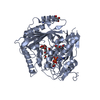
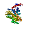
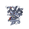
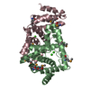
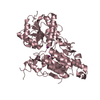
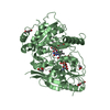
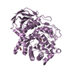
| Title | Crystal structure of TcCGT1 in complex with UDP | ||||||
 Components Components | C-glycosyltransferase | ||||||
 Keywords Keywords | TRANSFERASE / C-glycosyltransferase / glycosyltransferase | ||||||
| Function / homology |  Function and homology information Function and homology informationUDP-glycosyltransferase activity / Transferases; Glycosyltransferases; Hexosyltransferases / nucleotide binding Similarity search - Function | ||||||
| Biological species |  Trollius chinensis (jin lian hua) Trollius chinensis (jin lian hua) | ||||||
| Method |  X-RAY DIFFRACTION / SYNCHROTRON / Resolution: 1.85 Å X-RAY DIFFRACTION / SYNCHROTRON / Resolution: 1.85 Å | ||||||
 Authors Authors | Zhao, P. / Yun, C.H. | ||||||
 Citation Citation | Journal: Angew.Chem.Int.Ed.Engl. / Year: 2019 Title: Molecular and Structural Characterization of a Promiscuous C-Glycosyltransferase from Trollius chinensis. Authors: He, J.B. / Zhao, P. / Hu, Z.M. / Liu, S. / Kuang, Y. / Zhang, M. / Li, B. / Yun, C.H. / Qiao, X. / Ye, M. | ||||||
| History |
|
- Structure visualization
| Structure viewer | Molecule: MolmilJmol/JSmol |
|---|
- Downloads & links
-Download
| PDBx/mmCIF format | 6jtd.cif.gz | 219.7 KB | Display | PDBx/mmCIF format |
|---|---|---|---|---|
| PDB format | pdb6jtd.ent.gz | 173.5 KB | Display | PDB format |
| PDBx/mmJSON format | 6jtd.json.gz | Tree view | PDBx/mmJSON format | |
| Others |  Other downloads Other downloads |
-Validation report
| Arichive directory | https://data.pdbj.org/pub/pdb/validation_reports/jt/6jtdftp://data.pdbj.org/pub/pdb/validation_reports/jt/6jtd | HTTPS FTP |
|---|
-Related structure data
| Similar structure data |
|---|


-Links
 PDBj
PDBj


- Assembly
| Deposited unit | 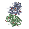
| ||||||||
|---|---|---|---|---|---|---|---|---|---|
| 1 | 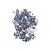
| ||||||||
| 2 | 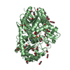
| ||||||||
| Unit cell |
|
-Components
| #1: Protein | Mass: 53953.070 Da / Num. of mol.: 2 Source method: isolated from a genetically manipulated source Source: (gene. exp.) Trollius chinensis (jin lian hua) / Production host:  #2: Chemical |   Type: RNA linking / Mass: 404.161 Da / Num. of mol.: 2 / Source method: obtained synthetically / Formula: C9H14N2O12P2 / Comment: UDP*YM Type: RNA linking / Mass: 404.161 Da / Num. of mol.: 2 / Source method: obtained synthetically / Formula: C9H14N2O12P2 / Comment: UDP*YM#3: Chemical | ChemComp-EDO /   Mass: 62.068 Da / Num. of mol.: 60 / Source method: obtained synthetically / Formula: C2H6O2 Mass: 62.068 Da / Num. of mol.: 60 / Source method: obtained synthetically / Formula: C2H6O2#4: Water | ChemComp-HOH / |  Mass: 18.015 Da / Num. of mol.: 675 / Source method: isolated from a natural source / Formula: H2O Mass: 18.015 Da / Num. of mol.: 675 / Source method: isolated from a natural source / Formula: H2O |
|---|
-Experimental details
-Experiment
| Experiment | Method: X-RAY DIFFRACTION / Number of used crystals: 1 |
|---|
- Sample preparation
| Crystal | Density Matthews: 2.44 Å3/Da / Density % sol: 49.69 % |
|---|---|
| Crystal grow | Temperature: 293 K / Method: vapor diffusion, hanging drop Details: 0.1M potassium chloride, 0.02M Tris 7.0, 20% w/v PEG 4000 |
-Data collection
| Diffraction | Mean temperature: 100 K / Serial crystal experiment: N |
|---|---|
| Diffraction source | Source: SYNCHROTRON / Site: SSRF  / Beamline: BL18U1 / Wavelength: 0.9793 Å / Beamline: BL18U1 / Wavelength: 0.9793 Å |
| Detector | Type: DECTRIS PILATUS3 6M / Detector: PIXEL / Date: Nov 4, 2018 |
| Radiation | Protocol: SINGLE WAVELENGTH / Monochromatic (M) / Laue (L): M / Scattering type: x-ray |
| Radiation wavelength | Wavelength: 0.9793 Å / Relative weight: 1 |
| Reflection | Resolution: 1.85→50 Å / Num. obs: 88344 / % possible obs: 98.9 % / Redundancy: 3.4 % / CC1/2: 0.981 / Net I/σ(I): 11.6 |
| Reflection shell | Resolution: 1.85→1.9 Å / Redundancy: 3.2 % / Mean I/σ(I) obs: 2 / CC1/2: 0.696 / % possible all: 97.9 |
- Processing
| Software |
| |||||||||||||||||||||||||||||||||||||||||||||||||||||||||||||||||||||||||||||||||||||||||||||||||||||||||||||||||||||||||||||||||||||||||||||||||||||||||||||||||||||||||||||||||||||||||||||||||||||||||||||||||||||||||
|---|---|---|---|---|---|---|---|---|---|---|---|---|---|---|---|---|---|---|---|---|---|---|---|---|---|---|---|---|---|---|---|---|---|---|---|---|---|---|---|---|---|---|---|---|---|---|---|---|---|---|---|---|---|---|---|---|---|---|---|---|---|---|---|---|---|---|---|---|---|---|---|---|---|---|---|---|---|---|---|---|---|---|---|---|---|---|---|---|---|---|---|---|---|---|---|---|---|---|---|---|---|---|---|---|---|---|---|---|---|---|---|---|---|---|---|---|---|---|---|---|---|---|---|---|---|---|---|---|---|---|---|---|---|---|---|---|---|---|---|---|---|---|---|---|---|---|---|---|---|---|---|---|---|---|---|---|---|---|---|---|---|---|---|---|---|---|---|---|---|---|---|---|---|---|---|---|---|---|---|---|---|---|---|---|---|---|---|---|---|---|---|---|---|---|---|---|---|---|---|---|---|---|---|---|---|---|---|---|---|---|---|---|---|---|---|---|---|---|
| Refinement | Resolution: 1.85→39.682 Å / SU ML: 0.19 / Cross valid method: FREE R-VALUE / σ(F): 1.36 / Phase error: 20.06
| |||||||||||||||||||||||||||||||||||||||||||||||||||||||||||||||||||||||||||||||||||||||||||||||||||||||||||||||||||||||||||||||||||||||||||||||||||||||||||||||||||||||||||||||||||||||||||||||||||||||||||||||||||||||||
| Solvent computation | Shrinkage radii: 0.9 Å / VDW probe radii: 1.11 Å | |||||||||||||||||||||||||||||||||||||||||||||||||||||||||||||||||||||||||||||||||||||||||||||||||||||||||||||||||||||||||||||||||||||||||||||||||||||||||||||||||||||||||||||||||||||||||||||||||||||||||||||||||||||||||
| Refinement step | Cycle: LAST / Resolution: 1.85→39.682 Å
| |||||||||||||||||||||||||||||||||||||||||||||||||||||||||||||||||||||||||||||||||||||||||||||||||||||||||||||||||||||||||||||||||||||||||||||||||||||||||||||||||||||||||||||||||||||||||||||||||||||||||||||||||||||||||
| Refine LS restraints |
| |||||||||||||||||||||||||||||||||||||||||||||||||||||||||||||||||||||||||||||||||||||||||||||||||||||||||||||||||||||||||||||||||||||||||||||||||||||||||||||||||||||||||||||||||||||||||||||||||||||||||||||||||||||||||
| LS refinement shell |
|
