 Movie
Movie Controller

[English] 日本語
 Yorodumi
- PDB-3mzo: Crystal structure of a HD-domain phosphohydrolase (lin2634) from ... -
+ Open data

- Basic information
| Entry | Database: PDB / ID: 3mzo | ||||||
|---|---|---|---|---|---|---|---|
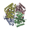
| Title | Crystal structure of a HD-domain phosphohydrolase (lin2634) from LISTERIA INNOCUA at 1.98 A resolution | ||||||
 Components Components | Lin2634 protein | ||||||
 Keywords Keywords | HYDROLASE / HD-domain phosphohydrolase / Structural Genomics / Joint Center for Structural Genomics / JCSG / Protein Structure Initiative / PSI-2 | ||||||
| Function / homology | 5'-deoxynucleotidase YfbR-like / Hypothetical protein af1432 / Hypothetical protein af1432 / Orthogonal Bundle / Mainly Alpha / metal ion binding / DI(HYDROXYETHYL)ETHER / Lin2634 protein Function and homology information Function and homology information | ||||||
| Biological species |  Listeria innocua (bacteria) Listeria innocua (bacteria) | ||||||
| Method |  X-RAY DIFFRACTION / SYNCHROTRON / MAD / Resolution: 1.98 Å X-RAY DIFFRACTION / SYNCHROTRON / MAD / Resolution: 1.98 Å | ||||||
 Authors Authors | Joint Center for Structural Genomics (JCSG) | ||||||
 Citation Citation | Journal: To be published Title: Crystal structure of a HD-domain phosphohydrolase (lin2634) from LISTERIA INNOCUA at 1.98 A resolution Authors: Joint Center for Structural Genomics (JCSG) | ||||||
| History |
|
- Structure visualization
| Structure viewer | Molecule: MolmilJmol/JSmol |
|---|
- Downloads & links
-Download
| PDBx/mmCIF format | 3mzo.cif.gz | 272.9 KB | Display | PDBx/mmCIF format |
|---|---|---|---|---|
| PDB format | pdb3mzo.ent.gz | 221.7 KB | Display | PDB format |
| PDBx/mmJSON format | 3mzo.json.gz | Tree view | PDBx/mmJSON format | |
| Others |  Other downloads Other downloads |
-Validation report
| Arichive directory | https://data.pdbj.org/pub/pdb/validation_reports/mz/3mzoftp://data.pdbj.org/pub/pdb/validation_reports/mz/3mzo | HTTPS FTP |
|---|
-Related structure data
| Similar structure data | |
|---|---|
| Other databases |


-Links
 PDBj
PDBj- Assembly
| Deposited unit | 
| ||||||||||||||||||||||||
|---|---|---|---|---|---|---|---|---|---|---|---|---|---|---|---|---|---|---|---|---|---|---|---|---|---|
| 1 | 
| ||||||||||||||||||||||||
| 2 | 
| ||||||||||||||||||||||||
| 3 | 
| ||||||||||||||||||||||||
| Unit cell |
| ||||||||||||||||||||||||
| Components on special symmetry positions |
| ||||||||||||||||||||||||
| Noncrystallographic symmetry (NCS) | NCS domain:
NCS domain segments: Component-ID: 1 / Ens-ID: 1 / Beg auth comp-ID: MSE / Beg label comp-ID: MSE / End auth comp-ID: THR / End label comp-ID: THR / Refine code: 4 / Auth seq-ID: 1 - 208 / Label seq-ID: 2 - 209
|
-Components
| #1: Protein | Mass: 25168.629 Da / Num. of mol.: 3 Source method: isolated from a genetically manipulated source Source: (gene. exp.) Listeria innocua (bacteria) / Gene: lin2634 / Plasmid: SpeedET / Production host: #2: Chemical | ChemComp-CA /   Mass: 40.078 Da / Num. of mol.: 11 / Source method: obtained synthetically / Formula: Ca Mass: 40.078 Da / Num. of mol.: 11 / Source method: obtained synthetically / Formula: Ca#3: Chemical |   Mass: 106.120 Da / Num. of mol.: 2 / Source method: obtained synthetically / Formula: C4H10O3 Mass: 106.120 Da / Num. of mol.: 2 / Source method: obtained synthetically / Formula: C4H10O3#4: Water | ChemComp-HOH / |  Mass: 18.015 Da / Num. of mol.: 375 / Source method: isolated from a natural source / Formula: H2O Mass: 18.015 Da / Num. of mol.: 375 / Source method: isolated from a natural source / Formula: H2OHas protein modification | Y | Sequence details | THE CONSTRUCT WAS EXPRESSED WITH AN N-TERMINAL PURIFICATION TAG MGSDKIHHHHHHENLYFQG. THE TAG WAS ...THE CONSTRUCT WAS EXPRESSED WITH AN N-TERMINAL PURIFICATI | |
|---|
-Experimental details
-Experiment
| Experiment | Method: X-RAY DIFFRACTION / Number of used crystals: 1 |
|---|
- Sample preparation
| Crystal | Density Matthews: 2.32 Å3/Da / Density % sol: 47.07 % |
|---|---|
| Crystal grow | Temperature: 277 K / Method: vapor diffusion, sitting drop / pH: 7.5 Details: 0.2000M calcium chloride, 28.0000% polyethylene glycol 400, 0.1M HEPES pH 7.5, 0.005M 2'-deoxyadenosine 5'-monophosphate (dAMP), NANODROP, VAPOR DIFFUSION, SITTING DROP, temperature 277K |
-Data collection
| Diffraction | Mean temperature: 100 K | ||||||||||||||||||||||||||||||||||||||||||||||||||||||||||||||||||
|---|---|---|---|---|---|---|---|---|---|---|---|---|---|---|---|---|---|---|---|---|---|---|---|---|---|---|---|---|---|---|---|---|---|---|---|---|---|---|---|---|---|---|---|---|---|---|---|---|---|---|---|---|---|---|---|---|---|---|---|---|---|---|---|---|---|---|---|
| Diffraction source | Source: SYNCHROTRON / Site: SSRL  / Beamline: BL9-2 / Wavelength: 0.91837,0.97939,0.97925 / Beamline: BL9-2 / Wavelength: 0.91837,0.97939,0.97925 | ||||||||||||||||||||||||||||||||||||||||||||||||||||||||||||||||||
| Detector | Type: MARMOSAIC 325 mm CCD / Detector: CCD / Date: Mar 11, 2010 / Details: Flat collimating mirror, toroid focusing mirror | ||||||||||||||||||||||||||||||||||||||||||||||||||||||||||||||||||
| Radiation | Monochromator: Double crystal monochromator / Protocol: MAD / Monochromatic (M) / Laue (L): M / Scattering type: x-ray | ||||||||||||||||||||||||||||||||||||||||||||||||||||||||||||||||||
| Radiation wavelength |
| ||||||||||||||||||||||||||||||||||||||||||||||||||||||||||||||||||
| Reflection | Resolution: 1.98→48.741 Å / Num. obs: 47810 / % possible obs: 99.4 % / Observed criterion σ(I): -3 / Biso Wilson estimate: 29.174 Å2 / Rmerge(I) obs: 0.053 / Net I/σ(I): 15.73 | ||||||||||||||||||||||||||||||||||||||||||||||||||||||||||||||||||
| Reflection shell |
|
-Phasing
| Phasing | Method: MAD |
|---|
- Processing
| Software |
| ||||||||||||||||||||||||||||||||||||||||||||||||||||||||||||||||||||||||||||||||||||||||||||||||||||
|---|---|---|---|---|---|---|---|---|---|---|---|---|---|---|---|---|---|---|---|---|---|---|---|---|---|---|---|---|---|---|---|---|---|---|---|---|---|---|---|---|---|---|---|---|---|---|---|---|---|---|---|---|---|---|---|---|---|---|---|---|---|---|---|---|---|---|---|---|---|---|---|---|---|---|---|---|---|---|---|---|---|---|---|---|---|---|---|---|---|---|---|---|---|---|---|---|---|---|---|---|---|
| Refinement | Method to determine structure: MAD / Resolution: 1.98→48.741 Å / Cor.coef. Fo:Fc: 0.966 / Cor.coef. Fo:Fc free: 0.953 / Occupancy max: 1 / Occupancy min: 0.3 / SU B: 6.819 / SU ML: 0.098 / Cross valid method: THROUGHOUT / σ(F): 0 / ESU R Free: 0.14 Stereochemistry target values: MAXIMUM LIKELIHOOD WITH PHASES Details: 1. HYDROGENS HAVE BEEN ADDED IN THE RIDING POSITIONS. 2. A MET-INHIBITION PROTOCOL WAS USED FOR SELENOMETHIONINE INCORPORATION DURING PROTEIN EXPRESSION. THE OCCUPANCY OF THE SE ATOMS IN THE ...Details: 1. HYDROGENS HAVE BEEN ADDED IN THE RIDING POSITIONS. 2. A MET-INHIBITION PROTOCOL WAS USED FOR SELENOMETHIONINE INCORPORATION DURING PROTEIN EXPRESSION. THE OCCUPANCY OF THE SE ATOMS IN THE MSE RESIDUES WAS REDUCED TO 0.75 FOR THE REDUCED SCATTERING POWER DUE TO PARTIAL S-MET INCORPORATION. 3. ATOM RECORD CONTAINS SUM OF TLS AND RESIDUAL B FACTORS. ANISOU RECORD CONTAINS SUM OF TLS AND RESIDUAL U FACTORS. 4. POLYETHYLENE GLYCOL (PEG), CALCIUM (CA) MODELED ARE PRESENT IN CRYSTALLIZATION/CRYO CANDITIONS.
| ||||||||||||||||||||||||||||||||||||||||||||||||||||||||||||||||||||||||||||||||||||||||||||||||||||
| Solvent computation | Ion probe radii: 0.8 Å / Shrinkage radii: 0.8 Å / VDW probe radii: 1.2 Å / Solvent model: MASK | ||||||||||||||||||||||||||||||||||||||||||||||||||||||||||||||||||||||||||||||||||||||||||||||||||||
| Displacement parameters | Biso max: 90.44 Å2 / Biso mean: 36.888 Å2 / Biso min: 5.38 Å2
| ||||||||||||||||||||||||||||||||||||||||||||||||||||||||||||||||||||||||||||||||||||||||||||||||||||
| Refinement step | Cycle: LAST / Resolution: 1.98→48.741 Å
| ||||||||||||||||||||||||||||||||||||||||||||||||||||||||||||||||||||||||||||||||||||||||||||||||||||
| Refine LS restraints |
| ||||||||||||||||||||||||||||||||||||||||||||||||||||||||||||||||||||||||||||||||||||||||||||||||||||
| Refine LS restraints NCS | Ens-ID: 1 / Number: 2445 / Refine-ID: X-RAY DIFFRACTION
| ||||||||||||||||||||||||||||||||||||||||||||||||||||||||||||||||||||||||||||||||||||||||||||||||||||
| LS refinement shell | Resolution: 1.98→2.031 Å / Total num. of bins used: 20
| ||||||||||||||||||||||||||||||||||||||||||||||||||||||||||||||||||||||||||||||||||||||||||||||||||||
| Refinement TLS params. | Method: refined / Refine-ID: X-RAY DIFFRACTION
| ||||||||||||||||||||||||||||||||||||||||||||||||||||||||||||||||||||||||||||||||||||||||||||||||||||
| Refinement TLS group |
|
